

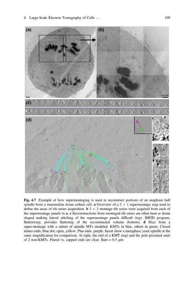
Fig. 4.7 Example of how supermontaging is used to reconstruct portions of an anaphase half
spindle from a mammalian tissue culture cell. a Overview of a 2 Â 1 supermontage map used to
define the areas of tilt series acquisition. b 3 Â 3 montage tilt series were acquired from each of
the supermontage panels in a. c Reconstructions from montaged tilt series are often bent or dome
shaped making lateral stitching of the supermontage panels difficult (top). IMOD program,
flattenwarp, provides flattening of the reconstructed volume (bottom). d Slice from a
super-montage with a subset of spindle MTs modeled. KMTs in blue, others in green. Closed
minus ends, blue dot; open, yellow. Plus ends, purple. Insert show a metaphase yeast spindle at the
same magnification for comparison. At right, the end of a KMT (top) and the pole-proximal ends
of 2 non-KMTs. Flared vs. capped ends are clear. Bars = 0.5 µm
4 Large-Scale Electron Tomography of Cells …
109
spindle from a mammalian tissue culture cell. a Overview of a 2 Â 1 supermontage map used to
define the areas of tilt series acquisition. b 3 Â 3 montage tilt series were acquired from each of
the supermontage panels in a. c Reconstructions from montaged tilt series are often bent or dome
shaped making lateral stitching of the supermontage panels difficult (top). IMOD program,
flattenwarp, provides flattening of the reconstructed volume (bottom). d Slice from a
super-montage with a subset of spindle MTs modeled. KMTs in blue, others in green. Closed
minus ends, blue dot; open, yellow. Plus ends, purple. Insert show a metaphase yeast spindle at the
same magnification for comparison. At right, the end of a KMT (top) and the pole-proximal ends
of 2 non-KMTs. Flared vs. capped ends are clear. Bars = 0.5 µm
4 Large-Scale Electron Tomography of Cells …
109