

reconstruction. Recent advances in the SerialEM program provide automated
acquisition from multiple positions, including automated serial section tilt series
and supermontage acquisition. The IMOD package provides alignment methods
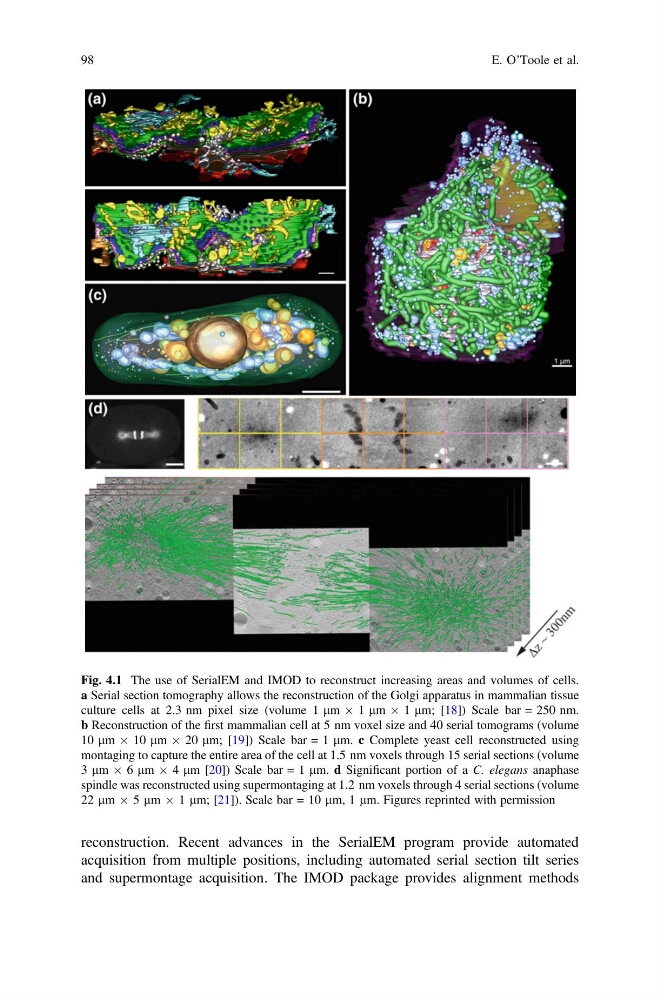
Fig. 4.1 The use of SerialEM and IMOD to reconstruct increasing areas and volumes of cells.
a Serial section tomography allows the reconstruction of the Golgi apparatus in mammalian tissue
culture cells at 2.3 nm pixel size (volume 1 lm  1 lm  1 lm; [18]) Scale bar = 250 nm.
b Reconstruction of the first mammalian cell at 5 nm voxel size and 40 serial tomograms (volume
10 lm  10 lm  20 lm; [19]) Scale bar = 1 lm. c Complete yeast cell reconstructed using
montaging to capture the entire area of the cell at 1.5 nm voxels through 15 serial sections (volume
3 lm  6 lm  4 lm [20]) Scale bar = 1 lm. d Significant portion of a C. elegans anaphase
spindle was reconstructed using supermontaging at 1.2 nm voxels through 4 serial sections (volume
22 lm  5 lm  1 lm; [21]). Scale bar = 10 lm, 1 lm. Figures reprinted with permission
98
E. O’Toole et al.
acquisition from multiple positions, including automated serial section tilt series
and supermontage acquisition. The IMOD package provides alignment methods
Fig. 4.1 The use of SerialEM and IMOD to reconstruct increasing areas and volumes of cells.
a Serial section tomography allows the reconstruction of the Golgi apparatus in mammalian tissue
culture cells at 2.3 nm pixel size (volume 1 lm  1 lm  1 lm; [18]) Scale bar = 250 nm.
b Reconstruction of the first mammalian cell at 5 nm voxel size and 40 serial tomograms (volume
10 lm  10 lm  20 lm; [19]) Scale bar = 1 lm. c Complete yeast cell reconstructed using
montaging to capture the entire area of the cell at 1.5 nm voxels through 15 serial sections (volume
3 lm  6 lm  4 lm [20]) Scale bar = 1 lm. d Significant portion of a C. elegans anaphase
spindle was reconstructed using supermontaging at 1.2 nm voxels through 4 serial sections (volume
22 lm  5 lm  1 lm; [21]). Scale bar = 10 lm, 1 lm. Figures reprinted with permission
98
E. O’Toole et al.