

Fundamental to cell shape is peptidoglycan, a single giant molecule of interlinked glycans and peptides that confers bacterial shape and mechanical protection,
and serves as a scaffold for cell division. Understanding the architecture of peptidoglycan is therefore crucial to understanding these processes, but its size and lack
of crystalline structure proved challenging for structural study by traditional techniques. ECT was uniquely able, therefore, to determine the architecture of both
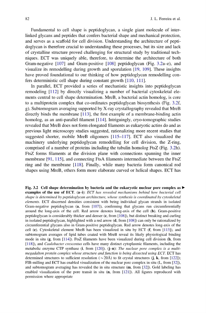
Gram-negative [107] and Gram-positive [108] peptidoglycan (Fig. 3.2a–e), and
visualize its remodelling during growth and sporulation [19, 109]. These insights
have proved foundational to our thinking of how peptidoglycan remodelling confers deterministic cell shape during constant growth [110, 111].
In parallel, ECT provided a series of mechanistic insights into peptidoglycan
remodeling [112] by directly visualizing a number of bacterial cytoskeletal elements central to cell shape determination. MreB, a bacterial actin homolog, is core
to a multiprotein complex that co-ordinates peptidoglycan biosynthesis (Fig. 3.2f,
g). Subtomogram averaging supported by X-ray crystallography revealed that MreB
directly binds the membrane [113], the first example of a membrane-binding actin
homolog, as an anti-parallel filament [114]. Intriguingly, cryo-tomographic studies
revealed that MreB does not form elongated filaments as eukaryotic actins do and as
previous light microscopy studies suggested, rationalizing more recent studies that
suggested shorter, mobile MreB oligomers [115–117]. ECT also visualized the
machinery underlying peptidoglycan remodelling for cell division, the Z-ring,
comprised of a number of proteins including the tubulin homolog FtsZ (Fig. 3.2h).
FtsZ forms filaments at the division plane with connections spanning the inner
membrane [91, 115], and connecting FtsA filaments intermediate between the FtsZ
ring and the membrane [118]. Finally, while many bacteria form canonical rod
shapes using MreB, others form more elaborate curved or helical shapes. ECT has
c
Fig. 3.2 Cell shape determination by bacteria and the eukaryotic nuclear pore complex as
examples of the use of ECT. (a–i): ECT has revealed mechanisms behind how bacterial cell
shape is determined by peptidoglycan architecture, whose synthesis is coordinated by cytoskeletal
elements. ECT discerned densities consistent with being individual glycan strands in isolated
Gram-negative peptidoglycan (a, from [107]), confirming that glycans run circumferentially
around the long-axis of the cell. Red arrow denotes long-axis of the cell (b). Gram-positive
peptidoglycan is considerably thicker and denser (c, from [108]), but distinct breaking and curling
in isolated peptidoglycan, highlighted with a red arrow (d, from [108]) can only be rationalized by
circumferential glycans also in Gram-positive peptidoglycan. Red arrow denotes long-axis of the
cell (e). Cytoskeletal element MreB has been visualized in situ by ECT (f, from [113]), and
subtomogram averages of lipid tubes coated with MreB reveal its likely physiological binding
mode in situ (g, from [114]). FtsZ filaments have been visualized during cell division (h, from
[118]), and Caulobacter crescentus cells have many distinct cytoplasmic filaments, including the
metabolic enzyme CTP synthase (i, from [120]). (j–n): The nuclear pore complex is a multimegadalton protein complex whose structure and function is being dissected using ECT. ECT has
determined structures to sufficient resolution (*20Å) to fit crystal structures (j, k, from [122]);
FIB-milling and ECT has enabled visualization of the nuclear pore complex in situ (L, from [32]),
and subtomogram averaging has revealed the in situ structure (m, from [32]). Gold labeling has
enabled visualization of the pore transit in situ (n, from [121]). All figures reproduced with
permission where appropriate
82
J. L. Ferreira et al.
and serves as a scaffold for cell division. Understanding the architecture of peptidoglycan is therefore crucial to understanding these processes, but its size and lack
of crystalline structure proved challenging for structural study by traditional techniques. ECT was uniquely able, therefore, to determine the architecture of both
Gram-negative [107] and Gram-positive [108] peptidoglycan (Fig. 3.2a–e), and
visualize its remodelling during growth and sporulation [19, 109]. These insights
have proved foundational to our thinking of how peptidoglycan remodelling confers deterministic cell shape during constant growth [110, 111].
In parallel, ECT provided a series of mechanistic insights into peptidoglycan
remodeling [112] by directly visualizing a number of bacterial cytoskeletal elements central to cell shape determination. MreB, a bacterial actin homolog, is core
to a multiprotein complex that co-ordinates peptidoglycan biosynthesis (Fig. 3.2f,
g). Subtomogram averaging supported by X-ray crystallography revealed that MreB
directly binds the membrane [113], the first example of a membrane-binding actin
homolog, as an anti-parallel filament [114]. Intriguingly, cryo-tomographic studies
revealed that MreB does not form elongated filaments as eukaryotic actins do and as
previous light microscopy studies suggested, rationalizing more recent studies that
suggested shorter, mobile MreB oligomers [115–117]. ECT also visualized the
machinery underlying peptidoglycan remodelling for cell division, the Z-ring,
comprised of a number of proteins including the tubulin homolog FtsZ (Fig. 3.2h).
FtsZ forms filaments at the division plane with connections spanning the inner
membrane [91, 115], and connecting FtsA filaments intermediate between the FtsZ
ring and the membrane [118]. Finally, while many bacteria form canonical rod
shapes using MreB, others form more elaborate curved or helical shapes. ECT has
c
Fig. 3.2 Cell shape determination by bacteria and the eukaryotic nuclear pore complex as
examples of the use of ECT. (a–i): ECT has revealed mechanisms behind how bacterial cell
shape is determined by peptidoglycan architecture, whose synthesis is coordinated by cytoskeletal
elements. ECT discerned densities consistent with being individual glycan strands in isolated
Gram-negative peptidoglycan (a, from [107]), confirming that glycans run circumferentially
around the long-axis of the cell. Red arrow denotes long-axis of the cell (b). Gram-positive
peptidoglycan is considerably thicker and denser (c, from [108]), but distinct breaking and curling
in isolated peptidoglycan, highlighted with a red arrow (d, from [108]) can only be rationalized by
circumferential glycans also in Gram-positive peptidoglycan. Red arrow denotes long-axis of the
cell (e). Cytoskeletal element MreB has been visualized in situ by ECT (f, from [113]), and
subtomogram averages of lipid tubes coated with MreB reveal its likely physiological binding
mode in situ (g, from [114]). FtsZ filaments have been visualized during cell division (h, from
[118]), and Caulobacter crescentus cells have many distinct cytoplasmic filaments, including the
metabolic enzyme CTP synthase (i, from [120]). (j–n): The nuclear pore complex is a multimegadalton protein complex whose structure and function is being dissected using ECT. ECT has
determined structures to sufficient resolution (*20Å) to fit crystal structures (j, k, from [122]);
FIB-milling and ECT has enabled visualization of the nuclear pore complex in situ (L, from [32]),
and subtomogram averaging has revealed the in situ structure (m, from [32]). Gold labeling has
enabled visualization of the pore transit in situ (n, from [121]). All figures reproduced with
permission where appropriate
82
J. L. Ferreira et al.