

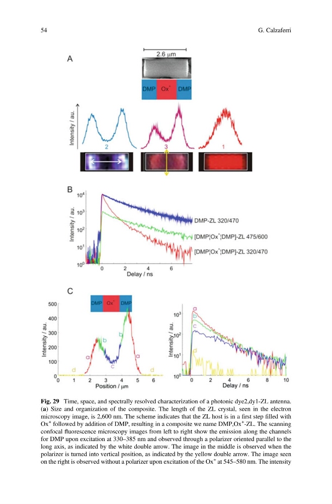
Fig. 29 Time, space, and spectrally resolved characterization of a photonic dye2,dy1-ZL antenna.
(a) Size and organization of the composite. The length of the ZL crystal, seen in the electron
microscopy image, is 2,600 nm. The scheme indicates that the ZL host is in a first step filled with
Ox
+ followed by addition of DMP, resulting in a composite we name DMP,Ox
+ -ZL. The scanning
confocal fluorescence microscopy images from left to right show the emission along the channels
for DMP upon excitation at 330–385 nm and observed through a polarizer oriented parallel to the
long axis, as indicated by the white double arrow. The image in the middle is observed when the
polarizer is turned into vertical position, as indicated by the yellow double arrow. The image seen
on the right is observed without a polarizer upon excitation of the Ox
+ at 545–580 nm. The intensity
54
G. Calzaferri
(a) Size and organization of the composite. The length of the ZL crystal, seen in the electron
microscopy image, is 2,600 nm. The scheme indicates that the ZL host is in a first step filled with
Ox
+ followed by addition of DMP, resulting in a composite we name DMP,Ox
+ -ZL. The scanning
confocal fluorescence microscopy images from left to right show the emission along the channels
for DMP upon excitation at 330–385 nm and observed through a polarizer oriented parallel to the
long axis, as indicated by the white double arrow. The image in the middle is observed when the
polarizer is turned into vertical position, as indicated by the yellow double arrow. The image seen
on the right is observed without a polarizer upon excitation of the Ox
+ at 545–580 nm. The intensity
54
G. Calzaferri