

[167, 168]. Absorption spectra of the S 0 –S 2 band have been measured on oriented
MeAcr
+ -ZL monolayers [128]. These data allow to interpret the single-crystal
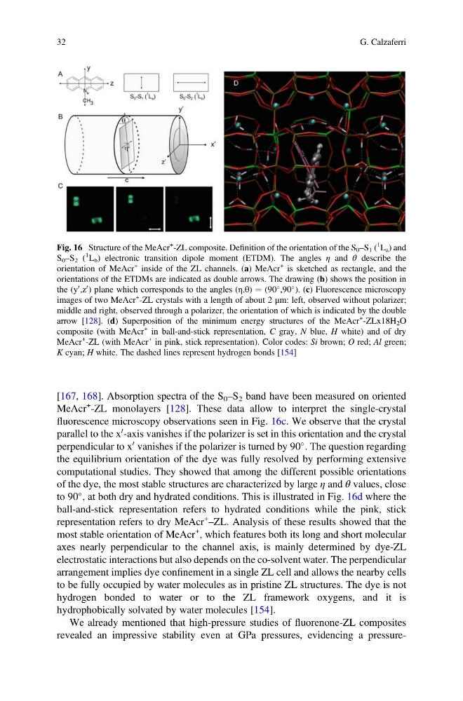
fluorescence microscopy observations seen in Fig. 16c. We observe that the crystal
parallel to the x
0 -axis vanishes if the polarizer is set in this orientation and the crystal
perpendicular to x
0 vanishes if the polarizer is turned by 90
. The question regarding
the equilibrium orientation of the dye was fully resolved by performing extensive
computational studies. They showed that among the different possible orientations
of the dye, the most stable structures are characterized by large η and θ values, close
to 90
, at both dry and hydrated conditions. This is illustrated in Fig. 16d where the
ball-and-stick representation refers to hydrated conditions while the pink, stick
representation refers to dry MeAcr
+
–ZL. Analysis of these results showed that the
most stable orientation of MeAcr
+ , which features both its long and short molecular
axes nearly perpendicular to the channel axis, is mainly determined by dye-ZL
electrostatic interactions but also depends on the co-solvent water. The perpendicular
arrangement implies dye confinement in a single ZL cell and allows the nearby cells
to be fully occupied by water molecules as in pristine ZL structures. The dye is not
hydrogen bonded to water or to the ZL framework oxygens, and it is
hydrophobically solvated by water molecules [154].
We already mentioned that high-pressure studies of fluorenone-ZL composites
revealed an impressive stability even at GPa pressures, evidencing a pressureFig. 16 Structure of the MeAcr
+ -ZL composite. Definition of the orientation of the S 0 –S 1 (
1
L a ) and
S 0 –S 2 (
1
L b ) electronic transition dipole moment (ETDM). The angles η and θ describe the
orientation of MeAcr
+ inside of the ZL channels. (a) MeAcr
+ is sketched as rectangle, and the
orientations of the ETDMs are indicated as double arrows. The drawing (b) shows the position in
the (y
0 ,z
0 ) plane which corresponds to the angles (η,θ) ¼ (90
,90
). (c) Fluorescence microscopy
images of two MeAcr
+ -ZL crystals with a length of about 2 μm: left, observed without polarizer;
middle and right, observed through a polarizer, the orientation of which is indicated by the double
arrow [128]. (d) Superposition of the minimum energy structures of the MeAcr
+ -ZLx18H 2 O
composite (with MeAcr
+ in ball-and-stick representation, C gray, N blue, H white) and of dry
MeAcr
+ -ZL (with MeAcr
+ in pink, stick representation). Color codes: Si brown; O red; Al green;
K cyan; H white. The dashed lines represent hydrogen bonds [154]
32
G. Calzaferri
MeAcr
+ -ZL monolayers [128]. These data allow to interpret the single-crystal
fluorescence microscopy observations seen in Fig. 16c. We observe that the crystal
parallel to the x
0 -axis vanishes if the polarizer is set in this orientation and the crystal
perpendicular to x
0 vanishes if the polarizer is turned by 90
. The question regarding
the equilibrium orientation of the dye was fully resolved by performing extensive
computational studies. They showed that among the different possible orientations
of the dye, the most stable structures are characterized by large η and θ values, close
to 90
, at both dry and hydrated conditions. This is illustrated in Fig. 16d where the
ball-and-stick representation refers to hydrated conditions while the pink, stick
representation refers to dry MeAcr
+
–ZL. Analysis of these results showed that the
most stable orientation of MeAcr
+ , which features both its long and short molecular
axes nearly perpendicular to the channel axis, is mainly determined by dye-ZL
electrostatic interactions but also depends on the co-solvent water. The perpendicular
arrangement implies dye confinement in a single ZL cell and allows the nearby cells
to be fully occupied by water molecules as in pristine ZL structures. The dye is not
hydrogen bonded to water or to the ZL framework oxygens, and it is
hydrophobically solvated by water molecules [154].
We already mentioned that high-pressure studies of fluorenone-ZL composites
revealed an impressive stability even at GPa pressures, evidencing a pressureFig. 16 Structure of the MeAcr
+ -ZL composite. Definition of the orientation of the S 0 –S 1 (
1
L a ) and
S 0 –S 2 (
1
L b ) electronic transition dipole moment (ETDM). The angles η and θ describe the
orientation of MeAcr
+ inside of the ZL channels. (a) MeAcr
+ is sketched as rectangle, and the
orientations of the ETDMs are indicated as double arrows. The drawing (b) shows the position in
the (y
0 ,z
0 ) plane which corresponds to the angles (η,θ) ¼ (90
,90
). (c) Fluorescence microscopy
images of two MeAcr
+ -ZL crystals with a length of about 2 μm: left, observed without polarizer;
middle and right, observed through a polarizer, the orientation of which is indicated by the double
arrow [128]. (d) Superposition of the minimum energy structures of the MeAcr
+ -ZLx18H 2 O
composite (with MeAcr
+ in ball-and-stick representation, C gray, N blue, H white) and of dry
MeAcr
+ -ZL (with MeAcr
+ in pink, stick representation). Color codes: Si brown; O red; Al green;
K cyan; H white. The dashed lines represent hydrogen bonds [154]
32
G. Calzaferri