

[113] or by immersing petals of different flowers into a suspension containing
dye-ZL crystals, Fig. 12g [116]. Hashimoto et al. examined the preparation of
close-packed monolayers on large areas. They prepared unidirectional alignment
in the horizontal packing structures (parallel alignment), Fig. 12f, by employing
pre-grooved substrates [132]. Utilization of optical tweezer [146], optical assembly
of bio-hybrid micro-robots [147], or opto-mechanically assisted assembly of
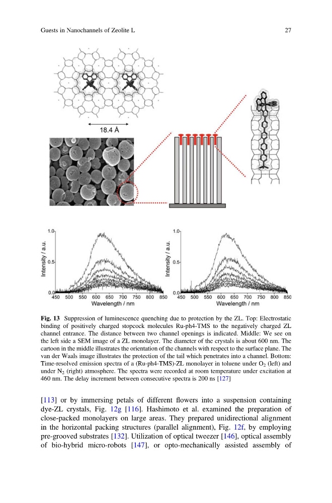
Fig. 13 Suppression of luminescence quenching due to protection by the ZL. Top: Electrostatic
binding of positively charged stopcock molecules Ru-ph4-TMS to the negatively charged ZL
channel entrance. The distance between two channel openings is indicated. Middle: We see on
the left side a SEM image of a ZL monolayer. The diameter of the crystals is about 600 nm. The
cartoon in the middle illustrates the orientation of the channels with respect to the surface plane. The
van der Waals image illustrates the protection of the tail which penetrates into a channel. Bottom:
Time-resolved emission spectra of a (Ru-ph4-TMS)-ZL monolayer in toluene under O 2 (left) and
under N 2 (right) atmosphere. The spectra were recorded at room temperature under excitation at
460 nm. The delay increment between consecutive spectra is 200 ns [127]
Guests in Nanochannels of Zeolite L
27
dye-ZL crystals, Fig. 12g [116]. Hashimoto et al. examined the preparation of
close-packed monolayers on large areas. They prepared unidirectional alignment
in the horizontal packing structures (parallel alignment), Fig. 12f, by employing
pre-grooved substrates [132]. Utilization of optical tweezer [146], optical assembly
of bio-hybrid micro-robots [147], or opto-mechanically assisted assembly of
Fig. 13 Suppression of luminescence quenching due to protection by the ZL. Top: Electrostatic
binding of positively charged stopcock molecules Ru-ph4-TMS to the negatively charged ZL
channel entrance. The distance between two channel openings is indicated. Middle: We see on
the left side a SEM image of a ZL monolayer. The diameter of the crystals is about 600 nm. The
cartoon in the middle illustrates the orientation of the channels with respect to the surface plane. The
van der Waals image illustrates the protection of the tail which penetrates into a channel. Bottom:
Time-resolved emission spectra of a (Ru-ph4-TMS)-ZL monolayer in toluene under O 2 (left) and
under N 2 (right) atmosphere. The spectra were recorded at room temperature under excitation at
460 nm. The delay increment between consecutive spectra is 200 ns [127]
Guests in Nanochannels of Zeolite L
27