

two magnitudes in sensitivity. Zhang et al. developed a faradaic aptasensor for
PDGF-BB and reached a LoD of 32 fM [45]. The lowest detection limit was
reached with 150 aM by He et al. [46] also using a faradaic impedance biosensor.
Already in 1986, Bard et al. [47] showed for recycling IDEs using redox
mediators that reducing the distance between the electrodes from 8 μm to 0.2 μm
increased the collection efficiency from 60% to 90%. Therefore, high expectations
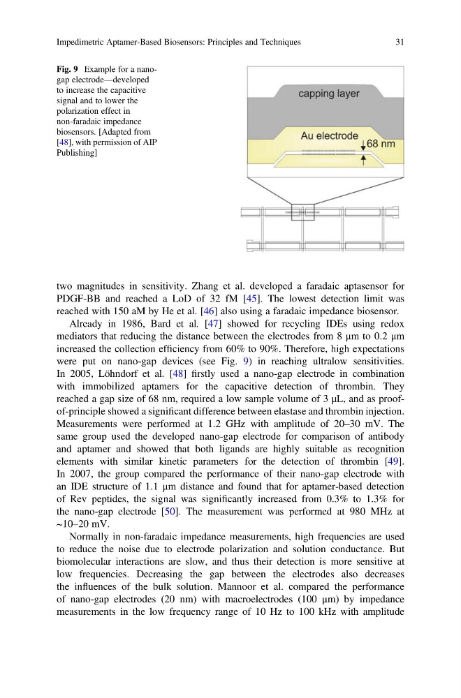
were put on nano-gap devices (see Fig. 9) in reaching ultralow sensitivities.
In 2005, Löhndorf et al. [48] firstly used a nano-gap electrode in combination
with immobilized aptamers for the capacitive detection of thrombin. They
reached a gap size of 68 nm, required a low sample volume of 3 μL, and as proofof-principle showed a significant difference between elastase and thrombin injection.
Measurements were performed at 1.2 GHz with amplitude of 20–30 mV. The
same group used the developed nano-gap electrode for comparison of antibody
and aptamer and showed that both ligands are highly suitable as recognition
elements with similar kinetic parameters for the detection of thrombin [49].
In 2007, the group compared the performance of their nano-gap electrode with
an IDE structure of 1.1 μm distance and found that for aptamer-based detection
of Rev peptides, the signal was significantly increased from 0.3% to 1.3% for
the nano-gap electrode [50]. The measurement was performed at 980 MHz at
~10–20 mV.
Normally in non-faradaic impedance measurements, high frequencies are used
to reduce the noise due to electrode polarization and solution conductance. But
biomolecular interactions are slow, and thus their detection is more sensitive at
low frequencies. Decreasing the gap between the electrodes also decreases
the influences of the bulk solution. Mannoor et al. compared the performance
of nano-gap electrodes (20 nm) with macroelectrodes (100 μm) by impedance
measurements in the low frequency range of 10 Hz to 100 kHz with amplitude
Fig. 9 Example for a nanogap electrode—developed
to increase the capacitive
signal and to lower the
polarization effect in
non-faradaic impedance
biosensors. [Adapted from
[48], with permission of AIP
Publishing]
Impedimetric Aptamer-Based Biosensors: Principles and Techniques
31
PDGF-BB and reached a LoD of 32 fM [45]. The lowest detection limit was
reached with 150 aM by He et al. [46] also using a faradaic impedance biosensor.
Already in 1986, Bard et al. [47] showed for recycling IDEs using redox
mediators that reducing the distance between the electrodes from 8 μm to 0.2 μm
increased the collection efficiency from 60% to 90%. Therefore, high expectations
were put on nano-gap devices (see Fig. 9) in reaching ultralow sensitivities.
In 2005, Löhndorf et al. [48] firstly used a nano-gap electrode in combination
with immobilized aptamers for the capacitive detection of thrombin. They
reached a gap size of 68 nm, required a low sample volume of 3 μL, and as proofof-principle showed a significant difference between elastase and thrombin injection.
Measurements were performed at 1.2 GHz with amplitude of 20–30 mV. The
same group used the developed nano-gap electrode for comparison of antibody
and aptamer and showed that both ligands are highly suitable as recognition
elements with similar kinetic parameters for the detection of thrombin [49].
In 2007, the group compared the performance of their nano-gap electrode with
an IDE structure of 1.1 μm distance and found that for aptamer-based detection
of Rev peptides, the signal was significantly increased from 0.3% to 1.3% for
the nano-gap electrode [50]. The measurement was performed at 980 MHz at
~10–20 mV.
Normally in non-faradaic impedance measurements, high frequencies are used
to reduce the noise due to electrode polarization and solution conductance. But
biomolecular interactions are slow, and thus their detection is more sensitive at
low frequencies. Decreasing the gap between the electrodes also decreases
the influences of the bulk solution. Mannoor et al. compared the performance
of nano-gap electrodes (20 nm) with macroelectrodes (100 μm) by impedance
measurements in the low frequency range of 10 Hz to 100 kHz with amplitude
Fig. 9 Example for a nanogap electrode—developed
to increase the capacitive
signal and to lower the
polarization effect in
non-faradaic impedance
biosensors. [Adapted from
[48], with permission of AIP
Publishing]
Impedimetric Aptamer-Based Biosensors: Principles and Techniques
31