

2.3 Application Range of the Selected Biophysical
Techniques
Different in vitro selection processes allow for the selection of aptamers for various
target classes, starting at aptamers against the smallest molecules, such as ions. Other
target classes for aptamer selection are small chemical molecules, peptides, nucleic
acids, proteins, high-molecular-weight protein complexes, particles, viruses, bacteria, and even whole cells. Due to the enormous differences between target classes in
size, charge, shape, and structure, there is currently no universally applicable
biophysical method for studying all possible aptamer-target combinations. The
limit of detection of the available biophysical methods is considerably influenced
by the rather small size of aptamers (low to middle kDa range) and the size of the
interaction partner (greatly varying from few Da as for ions to several MDa and more
for whole cells). Consequently, different biophysical methods have to be applied for
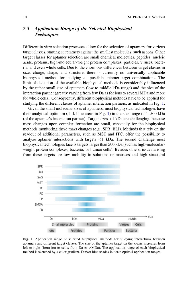
studying the different classes of aptamer interaction partners, as indicated in Fig. 1.
Given the small molecular sizes of aptamers, most biophysical technologies have
their analytical optimum (dark blue areas in Fig. 1) in the size range of 1–500 kDa
(of the aptamer’s interaction partner). Target sizes <1 kDa are challenging, because
mass changes upon complex formation are small, especially for the biophysical
methods monitoring these mass changes (e.g., SPR, BLI). Methods that rely on the
readout of additional parameters, such as MST and ITC, offer the possibility to
analyze aptamer interactions with targets <1 kDa. The second challenge most
biophysical technologies face is targets larger than 500 kDa (such as high-molecularweight protein complexes, bacteria, or human cells). Besides others, issues arising
from these targets are low mobility in solutions or matrices and high structural
Fig. 1 Application range of selected biophysical methods for studying interactions between
aptamers and different target classes. The size of the aptamer target on the x-axis increases from
left to right (from ion to cells; from Da to >MDa). The application range of each biophysical
method is sketched by a color gradient. Darker blue shades indicate optimal application ranges
10
M. Plach and T. Schubert
Techniques
Different in vitro selection processes allow for the selection of aptamers for various
target classes, starting at aptamers against the smallest molecules, such as ions. Other
target classes for aptamer selection are small chemical molecules, peptides, nucleic
acids, proteins, high-molecular-weight protein complexes, particles, viruses, bacteria, and even whole cells. Due to the enormous differences between target classes in
size, charge, shape, and structure, there is currently no universally applicable
biophysical method for studying all possible aptamer-target combinations. The
limit of detection of the available biophysical methods is considerably influenced
by the rather small size of aptamers (low to middle kDa range) and the size of the
interaction partner (greatly varying from few Da as for ions to several MDa and more
for whole cells). Consequently, different biophysical methods have to be applied for
studying the different classes of aptamer interaction partners, as indicated in Fig. 1.
Given the small molecular sizes of aptamers, most biophysical technologies have
their analytical optimum (dark blue areas in Fig. 1) in the size range of 1–500 kDa
(of the aptamer’s interaction partner). Target sizes <1 kDa are challenging, because
mass changes upon complex formation are small, especially for the biophysical
methods monitoring these mass changes (e.g., SPR, BLI). Methods that rely on the
readout of additional parameters, such as MST and ITC, offer the possibility to
analyze aptamer interactions with targets <1 kDa. The second challenge most
biophysical technologies face is targets larger than 500 kDa (such as high-molecularweight protein complexes, bacteria, or human cells). Besides others, issues arising
from these targets are low mobility in solutions or matrices and high structural
Fig. 1 Application range of selected biophysical methods for studying interactions between
aptamers and different target classes. The size of the aptamer target on the x-axis increases from
left to right (from ion to cells; from Da to >MDa). The application range of each biophysical
method is sketched by a color gradient. Darker blue shades indicate optimal application ranges
10
M. Plach and T. Schubert