

target binding. Thus this setup only works for aptamers which exhibit a large
conformational change upon target binding [52].
The methods for fluorescence biosensors listed above consist of labeled aptamers,
but there are some label-free methods as well. A competitive assay setup uses
aptamer-modified nanoparticles with free fluorophores intercalated to the aptamers.
Upon addition of the target, the fluorophores detach from the aptamer, and the
fluorescence signal increases, resulting in a signal-on sensor [53].
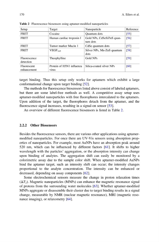
An overview of different fluorescence biosensors is listed in Table 2.
2.2.2 Other Biosensors
Besides the fluorescence sensors, there are various other applications using aptamermodified nanoparticles. For once there are UV-Vis sensors using absorption properties of nanoparticles. For example, most AuNPs have an absorption peak around
520 nm, which can be influenced by different factors [61]. It shifts to higher
wavelength with the particles’ aggregation, or the absorption intensity can change
upon binding of analytes. The aggregation shift can easily be monitored by a
colorimetric assay due to the sample color shift. When aptamer-modified AuNPs
bind the aptamer target, such an intensity shift can occur; the intensity changes
proportional to the analyte concentration. The intensity can be enhanced or
decreased, depending on assay components [62].
Some electrochemical sensors measure the change in proton relaxation times
(ΔT 2 ). Magnetic nanoparticles (MNPs) can enhance the magnetic resonance signal
of protons from the surrounding water molecules [63]. Whether aptamer-modified
MNPs aggregate or disassemble their cluster due to target binding results in a signal
change, measurable by NMR (nuclear magnetic resonance), MRI (magnetic resonance imaging), or relaxometry [64].
Table 2 Fluorescence biosensors using aptamer-modified nanoparticles
Setup
Target
Nanoparticle
Reference
FRET
Cocaine
Quantum dots
[55]
FRET
Human cardiac troponin I Gold NPs, CdSeS/ZnS quantum dots
[56]
FRET
Tumor marker Mucin 1
CdSe quantum dots
[57]
FRET
VEGF 165
Silver NPs, Mn-ZnS quantum
dots
[58]
Fluorescence
detection
Theophylline
Gold NPs
[59]
Fluorescent
enhancement
Protein of H5N1 influenza
virus
Silica-coated silver NPs
[60]
170
A. Eilers et al.
conformational change upon target binding [52].
The methods for fluorescence biosensors listed above consist of labeled aptamers,
but there are some label-free methods as well. A competitive assay setup uses
aptamer-modified nanoparticles with free fluorophores intercalated to the aptamers.
Upon addition of the target, the fluorophores detach from the aptamer, and the
fluorescence signal increases, resulting in a signal-on sensor [53].
An overview of different fluorescence biosensors is listed in Table 2.
2.2.2 Other Biosensors
Besides the fluorescence sensors, there are various other applications using aptamermodified nanoparticles. For once there are UV-Vis sensors using absorption properties of nanoparticles. For example, most AuNPs have an absorption peak around
520 nm, which can be influenced by different factors [61]. It shifts to higher
wavelength with the particles’ aggregation, or the absorption intensity can change
upon binding of analytes. The aggregation shift can easily be monitored by a
colorimetric assay due to the sample color shift. When aptamer-modified AuNPs
bind the aptamer target, such an intensity shift can occur; the intensity changes
proportional to the analyte concentration. The intensity can be enhanced or
decreased, depending on assay components [62].
Some electrochemical sensors measure the change in proton relaxation times
(ΔT 2 ). Magnetic nanoparticles (MNPs) can enhance the magnetic resonance signal
of protons from the surrounding water molecules [63]. Whether aptamer-modified
MNPs aggregate or disassemble their cluster due to target binding results in a signal
change, measurable by NMR (nuclear magnetic resonance), MRI (magnetic resonance imaging), or relaxometry [64].
Table 2 Fluorescence biosensors using aptamer-modified nanoparticles
Setup
Target
Nanoparticle
Reference
FRET
Cocaine
Quantum dots
[55]
FRET
Human cardiac troponin I Gold NPs, CdSeS/ZnS quantum dots
[56]
FRET
Tumor marker Mucin 1
CdSe quantum dots
[57]
FRET
VEGF 165
Silver NPs, Mn-ZnS quantum
dots
[58]
Fluorescence
detection
Theophylline
Gold NPs
[59]
Fluorescent
enhancement
Protein of H5N1 influenza
virus
Silica-coated silver NPs
[60]
170
A. Eilers et al.