

provide high molecular sensitivity (10
11
–10
12 mol/L) and independent location
depth of the emission source [56].
Moreover, image fusion techniques have been developed, and the combination of
CT and MRI with PET or SPECT modalities is now possible, offering a more
complete and accurate assessment of disease [94].
PET and SPECT substantially differ in the type of radionuclides used. PET is
based on the use of probes labeled with a radionuclide that decays by positron
emission. When the positron is combined with an electron, both are annihilated, and
their mass is converted. Each annihilation produces simultaneously two 511 keV
photons in opposite directions at an angle of 180
. SPECT radionuclides, such as
99m Tc,
111
In, and
67 Ga, decay with single photon emission. The use of both is
principally dependent on the radionuclide availability and imaging devices.
To create the radioactive probe, conjugation to radionuclides usually requires
labeling by isotopic or coordination reactions. This attachment creates the probe and
includes strict reaction time considerations due to radioactive decay and a very high
necessary final purity. Aptamers could be exposed to high-temperature reactions and
enable fast conjugation and high purity yields without altering their binding properties [11], a characteristic highly desired.
Both PET and SPECT are the most advanced molecular imaging modalities,
currently available in clinics. Besides their in vivo advantages, in addition to their
affinity and specificity, aptamers have great potential for this image modality in
which pharmacokinetics and tissue penetration play a central role [95]. The good
tissue penetration and fast clearance of aptamers lead to image acquisition in a
considerable time. Radioactive aptamer-based probes have been reported since
1997 [96]. Preclinical studies with aptamers in PET include the use of a positron
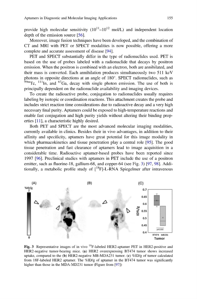
emitter, such as fluorine-18, gallium-68, and copper-64 (see Fig. 3) [97, 98]. Additionally, a metabolic profile study of [
18 F]-L-RNA Spiegelmer after intravenous
Fig. 3 Representative images of in vivo
18
F-labeled HER2-aptamer PET in HER2-positive and
HER2-negative tumor-bearing mice. (a) HER2 overexpressing BT474 tumor shows increased
uptake, compared to the (b) HER2-negative MB-MDA231 tumor. (c) %ID/g of tumor calculated
from 18F-labeled HER2 aptamer. The %ID/g of aptamer in the BT474 tumor was significantly
higher than those in the MDA-MD231 tumor (Figure from [97])
Aptamers in Diagnostic and Molecular Imaging Applications
155
11
–10
12 mol/L) and independent location
depth of the emission source [56].
Moreover, image fusion techniques have been developed, and the combination of
CT and MRI with PET or SPECT modalities is now possible, offering a more
complete and accurate assessment of disease [94].
PET and SPECT substantially differ in the type of radionuclides used. PET is
based on the use of probes labeled with a radionuclide that decays by positron
emission. When the positron is combined with an electron, both are annihilated, and
their mass is converted. Each annihilation produces simultaneously two 511 keV
photons in opposite directions at an angle of 180
. SPECT radionuclides, such as
99m Tc,
111
In, and
67 Ga, decay with single photon emission. The use of both is
principally dependent on the radionuclide availability and imaging devices.
To create the radioactive probe, conjugation to radionuclides usually requires
labeling by isotopic or coordination reactions. This attachment creates the probe and
includes strict reaction time considerations due to radioactive decay and a very high
necessary final purity. Aptamers could be exposed to high-temperature reactions and
enable fast conjugation and high purity yields without altering their binding properties [11], a characteristic highly desired.
Both PET and SPECT are the most advanced molecular imaging modalities,
currently available in clinics. Besides their in vivo advantages, in addition to their
affinity and specificity, aptamers have great potential for this image modality in
which pharmacokinetics and tissue penetration play a central role [95]. The good
tissue penetration and fast clearance of aptamers lead to image acquisition in a
considerable time. Radioactive aptamer-based probes have been reported since
1997 [96]. Preclinical studies with aptamers in PET include the use of a positron
emitter, such as fluorine-18, gallium-68, and copper-64 (see Fig. 3) [97, 98]. Additionally, a metabolic profile study of [
18 F]-L-RNA Spiegelmer after intravenous
Fig. 3 Representative images of in vivo
18
F-labeled HER2-aptamer PET in HER2-positive and
HER2-negative tumor-bearing mice. (a) HER2 overexpressing BT474 tumor shows increased
uptake, compared to the (b) HER2-negative MB-MDA231 tumor. (c) %ID/g of tumor calculated
from 18F-labeled HER2 aptamer. The %ID/g of aptamer in the BT474 tumor was significantly
higher than those in the MDA-MD231 tumor (Figure from [97])
Aptamers in Diagnostic and Molecular Imaging Applications
155