

dry ice/ethanol. Thawing can be done at room temperature or
higher (if it is desirable to be above a lipid phase-transition
temperature with, e.g., DPPC).
4. Transfer 1 mL of the suspension into the gastight syringes of a
LiposoFast extruder assemblage fitted with a 100 nm (or other
size) pore polycarbonate membrane. Push the lipid suspension
through the membrane 11–15 times on the day of use (see
Note 4).
5. To perform an LD experiment, if the analyte was not
incorporated at step 1, add the desired amount of peptide
and any probe molecule to the sample usually in a concentrated
solution (in a water miscible solvent) (see Notes 5 and 6).
Typically, one works with at least ~20:1 lipid:peptide ratios.
3.2 Flow LD Data
Collection
1. Liposome LD experiments require an instrument that produces
alternating beams of parallel and perpendicular light. Most CD
instruments now come with LD as a standard option. “Parallel”
is usually horizontal, which is what is needed for a Couette flow
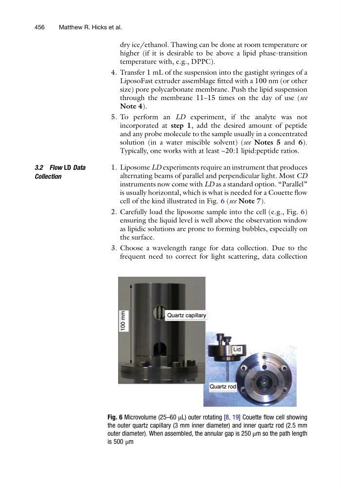
cell of the kind illustrated in Fig. 6 (see Note 7).
2. Carefully load the liposome sample into the cell (e.g., Fig. 6)
ensuring the liquid level is well above the observation window
as lipidic solutions are prone to forming bubbles, especially on
the surface.
3. Choose a wavelength range for data collection. Due to the
frequent need to correct for light scattering, data collection
Fig. 6 Microvolume (25–60 μL) outer rotating [8, 19] Couette flow cell showing
the outer quartz capillary (3 mm inner diameter) and inner quartz rod (2.5 mm
outer diameter). When assembled, the annular gap is 250 μm so the path length
is 500 μm
456
Matthew R. Hicks et al.
higher (if it is desirable to be above a lipid phase-transition
temperature with, e.g., DPPC).
4. Transfer 1 mL of the suspension into the gastight syringes of a
LiposoFast extruder assemblage fitted with a 100 nm (or other
size) pore polycarbonate membrane. Push the lipid suspension
through the membrane 11–15 times on the day of use (see
Note 4).
5. To perform an LD experiment, if the analyte was not
incorporated at step 1, add the desired amount of peptide
and any probe molecule to the sample usually in a concentrated
solution (in a water miscible solvent) (see Notes 5 and 6).
Typically, one works with at least ~20:1 lipid:peptide ratios.
3.2 Flow LD Data
Collection
1. Liposome LD experiments require an instrument that produces
alternating beams of parallel and perpendicular light. Most CD
instruments now come with LD as a standard option. “Parallel”
is usually horizontal, which is what is needed for a Couette flow
cell of the kind illustrated in Fig. 6 (see Note 7).
2. Carefully load the liposome sample into the cell (e.g., Fig. 6)
ensuring the liquid level is well above the observation window
as lipidic solutions are prone to forming bubbles, especially on
the surface.
3. Choose a wavelength range for data collection. Due to the
frequent need to correct for light scattering, data collection
Fig. 6 Microvolume (25–60 μL) outer rotating [8, 19] Couette flow cell showing
the outer quartz capillary (3 mm inner diameter) and inner quartz rod (2.5 mm
outer diameter). When assembled, the annular gap is 250 μm so the path length
is 500 μm
456
Matthew R. Hicks et al.