

3.14.1 Building an RNA
Chain Using RCrane
1. Start COOT.
2. Open the coordinates for the RNA–protein complex using
“File> Open Coordinates. . . .” from the COOT menu bar,
and select the appropriate PDB file.
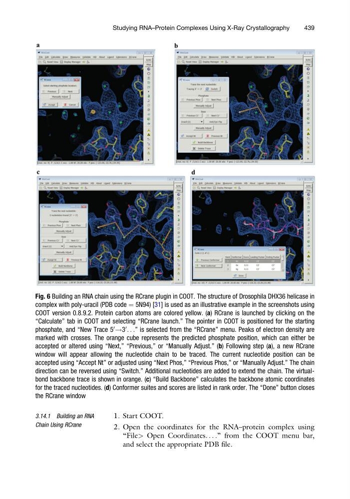
Fig. 6 Building an RNA chain using the RCrane plugin in COOT. The structure of Drosophila DHX36 helicase in
complex with poly-uracil (PDB code ¼ 5N94) [31] is used as an illustrative example in the screenshots using
COOT version 0.8.9.2. Protein carbon atoms are colored yellow. (a) RCrane is launched by clicking on the
“Calculate” tab in COOT and selecting “RCrane launch.” The pointer in COOT is positioned for the starting
phosphate, and “New Trace 5
0
!3
0
. . .” is selected from the “RCrane” menu. Peaks of electron density are
marked with crosses. The orange cube represents the predicted phosphate position, which can either be
accepted or altered using “Next,” “Previous,” or “Manually Adjust.” (b) Following step (a), a new RCrane
window will appear allowing the nucleotide chain to be traced. The current nucleotide position can be
accepted using “Accept Nt” or adjusted using “Next Phos,” “Previous Phos,” or “Manually Adjust.” The chain
direction can be reversed using “Switch.” Additional nucleotides are added to extend the chain. The virtualbond backbone trace is shown in orange. (c) “Build Backbone” calculates the backbone atomic coordinates
for the traced nucleotides. (d) Conformer suites and scores are listed in rank order. The “Done” button closes
the RCrane window
Studying RNA–Protein Complexes Using X-Ray Crystallography
439
Chain Using RCrane
1. Start COOT.
2. Open the coordinates for the RNA–protein complex using
“File> Open Coordinates. . . .” from the COOT menu bar,
and select the appropriate PDB file.
Fig. 6 Building an RNA chain using the RCrane plugin in COOT. The structure of Drosophila DHX36 helicase in
complex with poly-uracil (PDB code ¼ 5N94) [31] is used as an illustrative example in the screenshots using
COOT version 0.8.9.2. Protein carbon atoms are colored yellow. (a) RCrane is launched by clicking on the
“Calculate” tab in COOT and selecting “RCrane launch.” The pointer in COOT is positioned for the starting
phosphate, and “New Trace 5
0
!3
0
. . .” is selected from the “RCrane” menu. Peaks of electron density are
marked with crosses. The orange cube represents the predicted phosphate position, which can either be
accepted or altered using “Next,” “Previous,” or “Manually Adjust.” (b) Following step (a), a new RCrane
window will appear allowing the nucleotide chain to be traced. The current nucleotide position can be
accepted using “Accept Nt” or adjusted using “Next Phos,” “Previous Phos,” or “Manually Adjust.” The chain
direction can be reversed using “Switch.” Additional nucleotides are added to extend the chain. The virtualbond backbone trace is shown in orange. (c) “Build Backbone” calculates the backbone atomic coordinates
for the traced nucleotides. (d) Conformer suites and scores are listed in rank order. The “Done” button closes
the RCrane window
Studying RNA–Protein Complexes Using X-Ray Crystallography
439