

concentrations ranging between 1 and 20 mg/mL. 24-well Linbro
plates are commonly used to manually set up crystallization trials in
laboratories without access to a crystallization robot. For hanging
drop experiments, typically 0.5 or 1 μL of protein is mixed with an
equal volume of reservoir on a siliconized glass coverslip, which is
inverted and suspended over the reservoir solution. Alternatively,
sitting drop experiments can be performed using micro-bridges
(available from Hampton Research) placed in the Linbro plate,
which allow for much larger drop sizes to be used compared with
hanging drop experiments. The concentration difference between
the undersaturated drop and the reservoir at a higher precipitant
concentration results in vapor diffusion from the drop until the
solution concentration matches that of the reservoir. Consequently,
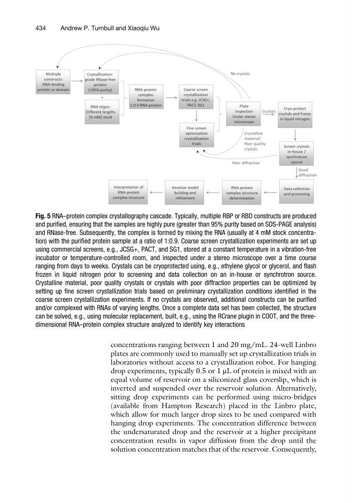
Fig. 5 RNA–protein complex crystallography cascade. Typically, multiple RBP or RBD constructs are produced
and purified, ensuring that the samples are highly pure (greater than 95% purity based on SDS-PAGE analysis)
and RNase-free. Subsequently, the complex is formed by mixing the RNA (usually at 4 mM stock concentration) with the purified protein sample at a ratio of 1:0.9. Coarse screen crystallization experiments are set up
using commercial screens, e.g., JCSG+, PACT, and SG1, stored at a constant temperature in a vibration-free
incubator or temperature-controlled room, and inspected under a stereo microscope over a time course
ranging from days to weeks. Crystals can be cryoprotected using, e.g., ethylene glycol or glycerol, and flash
frozen in liquid nitrogen prior to screening and data collection on an in-house or synchrotron source.
Crystalline material, poor quality crystals or crystals with poor diffraction properties can be optimized by
setting up fine screen crystallization trials based on preliminary crystallization conditions identified in the
coarse screen crystallization experiments. If no crystals are observed, additional constructs can be purified
and/or complexed with RNAs of varying lengths. Once a complete data set has been collected, the structure
can be solved, e.g., using molecular replacement, built, e.g., using the RCrane plugin in COOT, and the threedimensional RNA–protein complex structure analyzed to identify key interactions
434
Andrew P. Turnbull and Xiaoqiu Wu
plates are commonly used to manually set up crystallization trials in
laboratories without access to a crystallization robot. For hanging
drop experiments, typically 0.5 or 1 μL of protein is mixed with an
equal volume of reservoir on a siliconized glass coverslip, which is
inverted and suspended over the reservoir solution. Alternatively,
sitting drop experiments can be performed using micro-bridges
(available from Hampton Research) placed in the Linbro plate,
which allow for much larger drop sizes to be used compared with
hanging drop experiments. The concentration difference between
the undersaturated drop and the reservoir at a higher precipitant
concentration results in vapor diffusion from the drop until the
solution concentration matches that of the reservoir. Consequently,
Fig. 5 RNA–protein complex crystallography cascade. Typically, multiple RBP or RBD constructs are produced
and purified, ensuring that the samples are highly pure (greater than 95% purity based on SDS-PAGE analysis)
and RNase-free. Subsequently, the complex is formed by mixing the RNA (usually at 4 mM stock concentration) with the purified protein sample at a ratio of 1:0.9. Coarse screen crystallization experiments are set up
using commercial screens, e.g., JCSG+, PACT, and SG1, stored at a constant temperature in a vibration-free
incubator or temperature-controlled room, and inspected under a stereo microscope over a time course
ranging from days to weeks. Crystals can be cryoprotected using, e.g., ethylene glycol or glycerol, and flash
frozen in liquid nitrogen prior to screening and data collection on an in-house or synchrotron source.
Crystalline material, poor quality crystals or crystals with poor diffraction properties can be optimized by
setting up fine screen crystallization trials based on preliminary crystallization conditions identified in the
coarse screen crystallization experiments. If no crystals are observed, additional constructs can be purified
and/or complexed with RNAs of varying lengths. Once a complete data set has been collected, the structure
can be solved, e.g., using molecular replacement, built, e.g., using the RCrane plugin in COOT, and the threedimensional RNA–protein complex structure analyzed to identify key interactions
434
Andrew P. Turnbull and Xiaoqiu Wu