

autoradiography, fluorescence imaging, chemiluminescent imaging, and/or chromophore deposition, respectively. Radiolabeling
the nucleic acid at the 5
0 - or 3
0 -end with
32
P represents a widely
used, sensitive and inexpensive approach. RNA can also be covalently labeled with a fluorophore in labs where radioisotope use is
not permitted, and, with modern instrumentation, the detection of
fluorescent probes now rivals that of radioisotope-labeled RNAs. In
addition, unlabeled RNAs can be used and detected by postelectrophoretic staining with chromophores or fluorophores that
bind to the RNA. First, the RNA probe (see Note 5) is incubated
with a purified protein sample to initiate binding and formation of
the complex. The RNA–protein complex migrates through the gel
matrix more slowly than the free RNA probe, resulting in a migration shift relative to the non-bound RNA probe that can be
detected via the label or post-electrophoretic staining. The polyacrylamide or agarose gel composition and percentage may need to
be varied to optimize the separation of protein-bound and free
RNA. In addition, specificity can be determined using a competition reaction, where excess unlabeled RNA identical to the labeled
RNA is incubated in the binding reaction, resulting in a decrease in
the shifted signal when the labeled and unlabeled RNA sequences
compete for binding to the same protein. In order to use the
electrophoretic mobility shift assay to estimate dissociation constants for RNA–protein interactions, pure RBPs or RBDs are
required. Furthermore, the amount of free or protein-bound
RNA must be estimated by measuring the intensity of the bands,
e.g., in the autoradiograph or using fluorescence imaging. Gel
electrophoresis most likely affects the equilibrium between bound
and unbound RNA, hence dissociation constant values derived
from band shift assays are unlikely to represent absolute dissociation
constant values and are best used for comparative studies, e.g.,
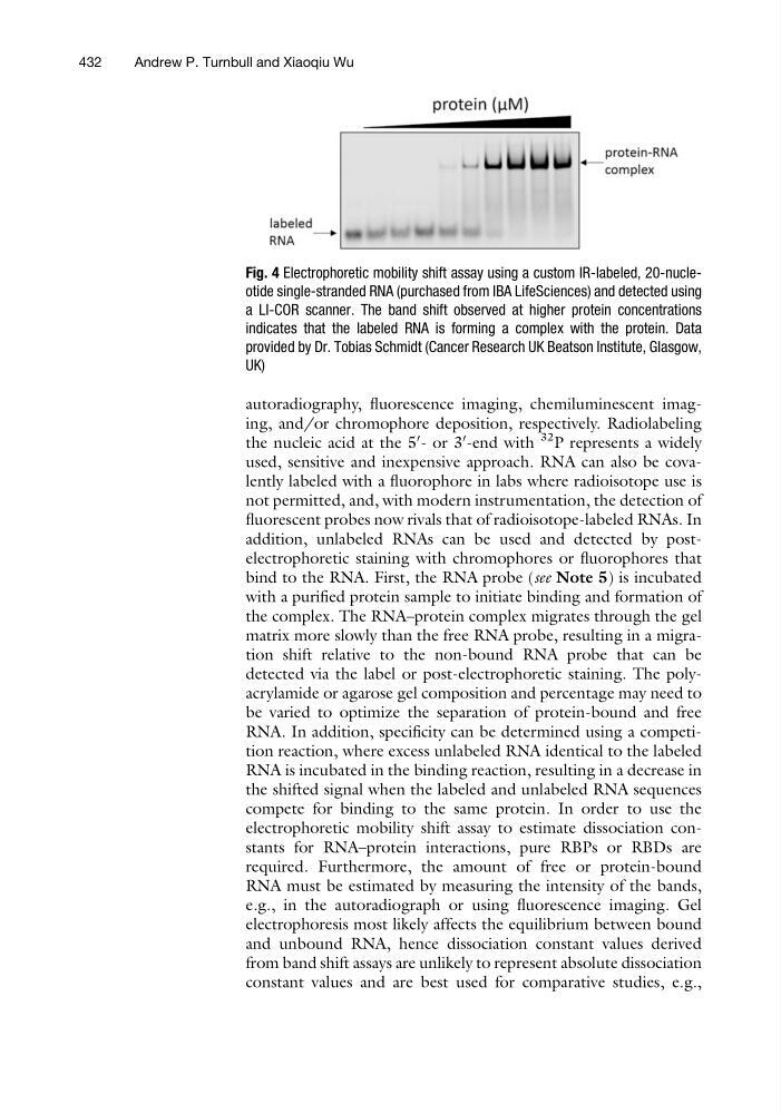
Fig. 4 Electrophoretic mobility shift assay using a custom IR-labeled, 20-nucleotide single-stranded RNA (purchased from IBA LifeSciences) and detected using
a LI-COR scanner. The band shift observed at higher protein concentrations
indicates that the labeled RNA is forming a complex with the protein. Data
provided by Dr. Tobias Schmidt (Cancer Research UK Beatson Institute, Glasgow,
UK)
432
Andrew P. Turnbull and Xiaoqiu Wu
the nucleic acid at the 5
0 - or 3
0 -end with
32
P represents a widely
used, sensitive and inexpensive approach. RNA can also be covalently labeled with a fluorophore in labs where radioisotope use is
not permitted, and, with modern instrumentation, the detection of
fluorescent probes now rivals that of radioisotope-labeled RNAs. In
addition, unlabeled RNAs can be used and detected by postelectrophoretic staining with chromophores or fluorophores that
bind to the RNA. First, the RNA probe (see Note 5) is incubated
with a purified protein sample to initiate binding and formation of
the complex. The RNA–protein complex migrates through the gel
matrix more slowly than the free RNA probe, resulting in a migration shift relative to the non-bound RNA probe that can be
detected via the label or post-electrophoretic staining. The polyacrylamide or agarose gel composition and percentage may need to
be varied to optimize the separation of protein-bound and free
RNA. In addition, specificity can be determined using a competition reaction, where excess unlabeled RNA identical to the labeled
RNA is incubated in the binding reaction, resulting in a decrease in
the shifted signal when the labeled and unlabeled RNA sequences
compete for binding to the same protein. In order to use the
electrophoretic mobility shift assay to estimate dissociation constants for RNA–protein interactions, pure RBPs or RBDs are
required. Furthermore, the amount of free or protein-bound
RNA must be estimated by measuring the intensity of the bands,
e.g., in the autoradiograph or using fluorescence imaging. Gel
electrophoresis most likely affects the equilibrium between bound
and unbound RNA, hence dissociation constant values derived
from band shift assays are unlikely to represent absolute dissociation
constant values and are best used for comparative studies, e.g.,
Fig. 4 Electrophoretic mobility shift assay using a custom IR-labeled, 20-nucleotide single-stranded RNA (purchased from IBA LifeSciences) and detected using
a LI-COR scanner. The band shift observed at higher protein concentrations
indicates that the labeled RNA is forming a complex with the protein. Data
provided by Dr. Tobias Schmidt (Cancer Research UK Beatson Institute, Glasgow,
UK)
432
Andrew P. Turnbull and Xiaoqiu Wu