

new classes of RBPs that mediate RNA interactions via disordered
regions, protein–protein interaction interfaces, and enzymatic cores
[1]. Determining the three-dimensional structures of RNA–protein complexes is critical to elucidate the relationship between
structure and biological function. X-ray crystallography represents
a powerful technique to determine the three-dimensional structures of RNA with unmodified full-length RBPs or RBDs at atomic
resolution. The first RNA–protein complex to be crystallized was E.
coli glutaminyl-tRNA synthetase in complex with its cognate tRNA
[2], which represented a major breakthrough in the field. Since
then (as of October 2020), 2971 RNA–protein complex structures
have been deposited in the RCSB Protein Data Bank (PDB) [3],
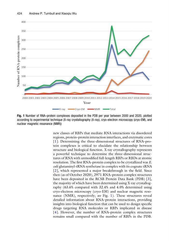
the majority of which have been determined using X-ray crystallography (63.6% compared with 32.4% and 4.0% determined using
cryo-electron microscopy (cryo-EM) and nuclear magnetic resonance (NMR), respectively; see Fig. 1). These structures reveal
detailed information about RNA–protein interactions, providing
insights into biological function that can be used to design specific
drugs targeting RNA molecules or RBPs implicated in disease
[4]. However, the number of RNA–protein complex structures
remains small compared with the number of RBPs in the PDB.
Fig. 1 Number of RNA–protein complexes deposited in the PDB per year between 2000 and 2020, plotted
according to experimental technique (X-ray crystallography (X-ray), cryo-electron microscopy (cryo-EM), and
nuclear magnetic resonance (NMR))
424
Andrew P. Turnbull and Xiaoqiu Wu
regions, protein–protein interaction interfaces, and enzymatic cores
[1]. Determining the three-dimensional structures of RNA–protein complexes is critical to elucidate the relationship between
structure and biological function. X-ray crystallography represents
a powerful technique to determine the three-dimensional structures of RNA with unmodified full-length RBPs or RBDs at atomic
resolution. The first RNA–protein complex to be crystallized was E.
coli glutaminyl-tRNA synthetase in complex with its cognate tRNA
[2], which represented a major breakthrough in the field. Since
then (as of October 2020), 2971 RNA–protein complex structures
have been deposited in the RCSB Protein Data Bank (PDB) [3],
the majority of which have been determined using X-ray crystallography (63.6% compared with 32.4% and 4.0% determined using
cryo-electron microscopy (cryo-EM) and nuclear magnetic resonance (NMR), respectively; see Fig. 1). These structures reveal
detailed information about RNA–protein interactions, providing
insights into biological function that can be used to design specific
drugs targeting RNA molecules or RBPs implicated in disease
[4]. However, the number of RNA–protein complex structures
remains small compared with the number of RBPs in the PDB.
Fig. 1 Number of RNA–protein complexes deposited in the PDB per year between 2000 and 2020, plotted
according to experimental technique (X-ray crystallography (X-ray), cryo-electron microscopy (cryo-EM), and
nuclear magnetic resonance (NMR))
424
Andrew P. Turnbull and Xiaoqiu Wu