

measuring analytes adsorption on a layer of actuated surface-bound
fluorescent probe, this combination of biophysical approaches can
be used for several applications, as binding kinetics and affinity,
protein diameter, conformational change, or even nuclease and
polymerase activity.
The originality of the switchSENSE
® technology lies in the
DNA strand bearing a fluorescent dye at one extremity, attached
on its opposite end to a gold-quenching surface. The complementary strand could be used alone or chemically coupled to an interaction partner (a protein). Hybridization of the DNA strands
generates a rigid negatively charged electro-switchable biosensor,
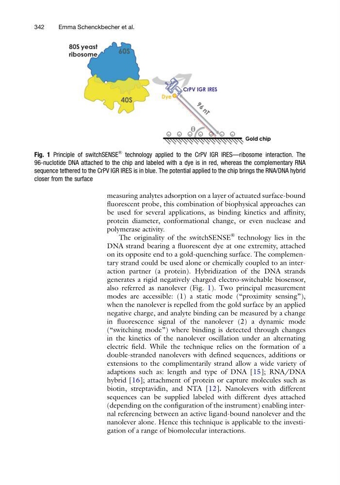
also referred as nanolever (Fig. 1). Two principal measurement
modes are accessible: (1) a static mode (“proximity sensing”),
when the nanolever is repelled from the gold surface by an applied
negative charge, and analyte binding can be measured by a change
in fluorescence signal of the nanolever (2) a dynamic mode
(“switching mode”) where binding is detected through changes
in the kinetics of the nanolever oscillation under an alternating
electric field. While the technique relies on the formation of a
double-stranded nanolevers with defined sequences, additions or
extensions to the complimentarily strand allow a wide variety of
adaptions such as: length and type of DNA [15]; RNA/DNA
hybrid [16]; attachment of protein or capture molecules such as
biotin, streptavidin, and NTA [12]. Nanolevers with different
sequences can be supplied labeled with different dyes attached
(depending on the configuration of the instrument) enabling internal referencing between an active ligand-bound nanolever and the
nanolever alone. Hence this technique is applicable to the investigation of a range of biomolecular interactions.
Fig. 1 Principle of switchSENSE
®
technology applied to the CrPV IGR IRES—ribosome interaction. The
96-nuclotide DNA attached to the chip and labeled with a dye is in red, whereas the complementary RNA
sequence tethered to the CrPV IGR IRES is in blue. The potential applied to the chip brings the RNA/DNA hybrid
closer from the surface
342
Emma Schenckbecher et al.
fluorescent probe, this combination of biophysical approaches can
be used for several applications, as binding kinetics and affinity,
protein diameter, conformational change, or even nuclease and
polymerase activity.
The originality of the switchSENSE
® technology lies in the
DNA strand bearing a fluorescent dye at one extremity, attached
on its opposite end to a gold-quenching surface. The complementary strand could be used alone or chemically coupled to an interaction partner (a protein). Hybridization of the DNA strands
generates a rigid negatively charged electro-switchable biosensor,
also referred as nanolever (Fig. 1). Two principal measurement
modes are accessible: (1) a static mode (“proximity sensing”),
when the nanolever is repelled from the gold surface by an applied
negative charge, and analyte binding can be measured by a change
in fluorescence signal of the nanolever (2) a dynamic mode
(“switching mode”) where binding is detected through changes
in the kinetics of the nanolever oscillation under an alternating
electric field. While the technique relies on the formation of a
double-stranded nanolevers with defined sequences, additions or
extensions to the complimentarily strand allow a wide variety of
adaptions such as: length and type of DNA [15]; RNA/DNA
hybrid [16]; attachment of protein or capture molecules such as
biotin, streptavidin, and NTA [12]. Nanolevers with different
sequences can be supplied labeled with different dyes attached
(depending on the configuration of the instrument) enabling internal referencing between an active ligand-bound nanolever and the
nanolever alone. Hence this technique is applicable to the investigation of a range of biomolecular interactions.
Fig. 1 Principle of switchSENSE
®
technology applied to the CrPV IGR IRES—ribosome interaction. The
96-nuclotide DNA attached to the chip and labeled with a dye is in red, whereas the complementary RNA
sequence tethered to the CrPV IGR IRES is in blue. The potential applied to the chip brings the RNA/DNA hybrid
closer from the surface
342
Emma Schenckbecher et al.