

DNase contamination. It is also possible to bake glassware and
tubes to remove RNases but this is not generally necessary.
2. Depending on the required resolution and size of macromolecules, the final acrylamide concentration should be adjusted
accordingly. We use 8% polyacrylamide for RNAs of ~10–50
bases. Larger proteins and longer nucleic acids should be
resolved on a lower percentage polyacrylamide gel. Smaller
substrates may resolve better on higher percentage gels.
3. Unpolymerized acrylamide is a potent neurotoxin and should
always be handled with caution. To minimize the risk of inhalation, we use a premade 40% acrylamide:bis-acrylamide (19:1
ratio) solution as a working stock and avoid powdered
5 0
1 5 0
1 0 0
2 0 0
2 5 0 3 0 0
3 5 0 4 0 0
4 5 0
5 0 0 1 ,0 0 0
2 ,0 0 0
a
b
6-FAM ‘blue’
labelled PRE RNA
Alexa647 ‘red’
labelled AU RNA
6-FAM ‘blue’
labelled PRE RNA
Alexa647 ‘red’
labelled AU RNA
100 nM probe
10 nM probe
0
spPuf3 (nM):
5 0
1 5 0
1 0 0
2 0 0
2 5 0 3 0 0
3 5 0 4 0 0
4 5 0
5 0 0 1 ,0 0 0
2 ,0 0 0
0
spPuf3 (nM):
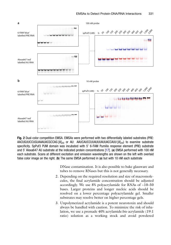
Fig. 2 Dual-color competition EMSA. EMSAs were performed with two differentially labeled substrates (PRE:
AACUGUUCCUGUAAAUACGCCAG [A] 30 or AU: AAUCAUCCUUAUUUAUUACCAUU [A] 30 ) to examine substrate
specificity. SpPuf3 PUM domain was incubated with 5
0
6-FAM Pumilio response element (PRE) substrate
and 5
0
Alexa647 AU substrate at the indicated protein concentrations [17]. (a) EMSA performed with 100 nM
each substrate. Scans at different excitation and emission wavelengths are shown on the left with overlaid
false color image on the right. (b) The same EMSA performed in (a) but with 10 nM each substrate
EMSAs to Detect Protein-DNA/RNA Interactions
331
tubes to remove RNases but this is not generally necessary.
2. Depending on the required resolution and size of macromolecules, the final acrylamide concentration should be adjusted
accordingly. We use 8% polyacrylamide for RNAs of ~10–50
bases. Larger proteins and longer nucleic acids should be
resolved on a lower percentage polyacrylamide gel. Smaller
substrates may resolve better on higher percentage gels.
3. Unpolymerized acrylamide is a potent neurotoxin and should
always be handled with caution. To minimize the risk of inhalation, we use a premade 40% acrylamide:bis-acrylamide (19:1
ratio) solution as a working stock and avoid powdered
5 0
1 5 0
1 0 0
2 0 0
2 5 0 3 0 0
3 5 0 4 0 0
4 5 0
5 0 0 1 ,0 0 0
2 ,0 0 0
a
b
6-FAM ‘blue’
labelled PRE RNA
Alexa647 ‘red’
labelled AU RNA
6-FAM ‘blue’
labelled PRE RNA
Alexa647 ‘red’
labelled AU RNA
100 nM probe
10 nM probe
0
spPuf3 (nM):
5 0
1 5 0
1 0 0
2 0 0
2 5 0 3 0 0
3 5 0 4 0 0
4 5 0
5 0 0 1 ,0 0 0
2 ,0 0 0
0
spPuf3 (nM):
Fig. 2 Dual-color competition EMSA. EMSAs were performed with two differentially labeled substrates (PRE:
AACUGUUCCUGUAAAUACGCCAG [A] 30 or AU: AAUCAUCCUUAUUUAUUACCAUU [A] 30 ) to examine substrate
specificity. SpPuf3 PUM domain was incubated with 5
0
6-FAM Pumilio response element (PRE) substrate
and 5
0
Alexa647 AU substrate at the indicated protein concentrations [17]. (a) EMSA performed with 100 nM
each substrate. Scans at different excitation and emission wavelengths are shown on the left with overlaid
false color image on the right. (b) The same EMSA performed in (a) but with 10 nM each substrate
EMSAs to Detect Protein-DNA/RNA Interactions
331