

the anode and the protein-bound nucleic acids are found “shifted”
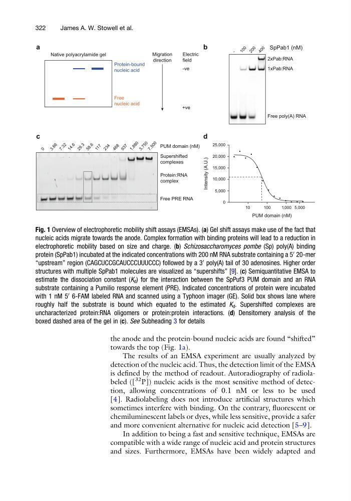
towards the top (Fig. 1a).
The results of an EMSA experiment are usually analyzed by
detection of the nucleic acid. Thus, the detection limit of the EMSA
is defined by the method of readout. Autoradiography of radiolabeled ([
32 P]) nucleic acids is the most sensitive method of detection, allowing concentrations of 0.1 nM or less to be used
[4]. Radiolabeling does not introduce artificial structures which
sometimes interfere with binding. On the contrary, fluorescent or
chemiluminescent labels or dyes, while less sensitive, provide a safer
and more convenient alternative for nucleic acid detection [5–9].
In addition to being a fast and sensitive technique, EMSAs are
compatible with a wide range of nucleic acid and protein structures
and sizes. Furthermore, EMSAs have been widely adapted and
3 . 6 6
1 4 . 6
7 . 3 2
2 9 . 3
5 8 . 6
1 1 7
2 3 4
a
4 6 8
9 3 7
1 , 8 8 0
3 , 7 5 0
7 , 5 0 0
PUM domain (nM)
Free PRE RNA
Protein:RNA
complex
Supershifted
complexes
b
10
100
1,000 5,000
0
5,000
10,000
15,000
20,000
25,000
Migration
direction
Electric
field
-ve
+ve
Free
nucleic acid
Protein-bound
nucleic acid
Native polyacrylamide gel
c
d
-
1 0 0 2 0 0 4 0 0
SpPab1 (nM)
Free poly(A) RNA
1xPab:RNA
2xPab:RNA
0
PUM domain (nM)
Intensity (A.U.)
Fig. 1 Overview of electrophoretic mobility shift assays (EMSAs). (a) Gel shift assays make use of the fact that
nucleic acids migrate towards the anode. Complex formation with binding proteins will lead to a reduction in
electrophoretic mobility based on size and charge. (b) Schizosaccharomyces pombe (Sp) poly(A) binding
protein (SpPab1) incubated at the indicated concentrations with 200 nM RNA substrate containing a 5
0
20-mer
“upstream” region (CAGCUCCGCAUCCCUUUCCC) followed by a 3
0
poly(A) tail of 30 adenosines. Higher order
structures with multiple SpPab1 molecules are visualized as “supershifts” [9]. (c) Semiquantitative EMSA to
estimate the dissociation constant (K d ) for the interaction between the SpPuf3 PUM domain and an RNA
substrate containing a Pumilio response element (PRE). Indicated concentrations of protein were incubated
with 1 nM 5
0
6-FAM labeled RNA and scanned using a Typhoon imager (GE). Solid box shows lane where
roughly half the substrate is bound which equated to the estimated K d . Supershifted complexes are
uncharacterized protein:RNA oligomers or protein:protein interactions. (d) Densitomery analysis of the
boxed dashed area of the gel in (c). See Subheading 3 for details
322
James A. W. Stowell et al.
towards the top (Fig. 1a).
The results of an EMSA experiment are usually analyzed by
detection of the nucleic acid. Thus, the detection limit of the EMSA
is defined by the method of readout. Autoradiography of radiolabeled ([
32 P]) nucleic acids is the most sensitive method of detection, allowing concentrations of 0.1 nM or less to be used
[4]. Radiolabeling does not introduce artificial structures which
sometimes interfere with binding. On the contrary, fluorescent or
chemiluminescent labels or dyes, while less sensitive, provide a safer
and more convenient alternative for nucleic acid detection [5–9].
In addition to being a fast and sensitive technique, EMSAs are
compatible with a wide range of nucleic acid and protein structures
and sizes. Furthermore, EMSAs have been widely adapted and
3 . 6 6
1 4 . 6
7 . 3 2
2 9 . 3
5 8 . 6
1 1 7
2 3 4
a
4 6 8
9 3 7
1 , 8 8 0
3 , 7 5 0
7 , 5 0 0
PUM domain (nM)
Free PRE RNA
Protein:RNA
complex
Supershifted
complexes
b
10
100
1,000 5,000
0
5,000
10,000
15,000
20,000
25,000
Migration
direction
Electric
field
-ve
+ve
Free
nucleic acid
Protein-bound
nucleic acid
Native polyacrylamide gel
c
d
-
1 0 0 2 0 0 4 0 0
SpPab1 (nM)
Free poly(A) RNA
1xPab:RNA
2xPab:RNA
0
PUM domain (nM)
Intensity (A.U.)
Fig. 1 Overview of electrophoretic mobility shift assays (EMSAs). (a) Gel shift assays make use of the fact that
nucleic acids migrate towards the anode. Complex formation with binding proteins will lead to a reduction in
electrophoretic mobility based on size and charge. (b) Schizosaccharomyces pombe (Sp) poly(A) binding
protein (SpPab1) incubated at the indicated concentrations with 200 nM RNA substrate containing a 5
0
20-mer
“upstream” region (CAGCUCCGCAUCCCUUUCCC) followed by a 3
0
poly(A) tail of 30 adenosines. Higher order
structures with multiple SpPab1 molecules are visualized as “supershifts” [9]. (c) Semiquantitative EMSA to
estimate the dissociation constant (K d ) for the interaction between the SpPuf3 PUM domain and an RNA
substrate containing a Pumilio response element (PRE). Indicated concentrations of protein were incubated
with 1 nM 5
0
6-FAM labeled RNA and scanned using a Typhoon imager (GE). Solid box shows lane where
roughly half the substrate is bound which equated to the estimated K d . Supershifted complexes are
uncharacterized protein:RNA oligomers or protein:protein interactions. (d) Densitomery analysis of the
boxed dashed area of the gel in (c). See Subheading 3 for details
322
James A. W. Stowell et al.