

responds very sensitively to properties of its local microenvironment. Thermal unfolding is measured by following the intrinsic Trp
steady-state fluorescence intensity and the position of the emission
maximum [27, 28]. The ratio of the fluorescence intensities at two
different wavelengths, 330 nm (folded) and 350 nm (unfolded), is
used to follow the change in the environment of Trp residues
during temperature-dependent unfolding of the protein. This
ratio shows a sharp increase of the signal near the midpoint of
thermal unfolding. T m can be calculated from the maximum of
the first derivative. The applicability of DSF of intrinsic fluorophores necessarily requires the presence of Trp (or high levels of
Tyr) in the folded core of the protein that will be exposed upon
unfolding. Moreover, it is necessary to exclude (e.g., with complementary DLS experiments) the possibility that observed signal
changes are caused by any aggregation as this will also lead to
changes in the fluorophore environment. A comprehensive understanding of the measurement, advantages, and drawbacks can be
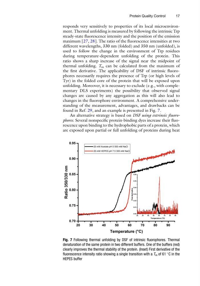
found in Ref. 29, and an example is presented in Fig. 7.
An alternative strategy is based on DSF using extrinsic fluorophores. Several nonspecific protein-binding dyes increase their fluorescence upon binding to the hydrophobic parts of a protein, which
are exposed upon partial or full unfolding of proteins during heat
Fig. 7 Following thermal unfolding by DSF of intrinsic fluorophores. Thermal
denaturation of the same protein in two different buffers. One of the buffers (red)
clearly improves the thermal stability of the protein. (Inset) First derivative of the
fluorescence intensity ratio showing a single transition with a T m of 61
C in the
HEPES buffer
Protein Quality Control
17
steady-state fluorescence intensity and the position of the emission
maximum [27, 28]. The ratio of the fluorescence intensities at two
different wavelengths, 330 nm (folded) and 350 nm (unfolded), is
used to follow the change in the environment of Trp residues
during temperature-dependent unfolding of the protein. This
ratio shows a sharp increase of the signal near the midpoint of
thermal unfolding. T m can be calculated from the maximum of
the first derivative. The applicability of DSF of intrinsic fluorophores necessarily requires the presence of Trp (or high levels of
Tyr) in the folded core of the protein that will be exposed upon
unfolding. Moreover, it is necessary to exclude (e.g., with complementary DLS experiments) the possibility that observed signal
changes are caused by any aggregation as this will also lead to
changes in the fluorophore environment. A comprehensive understanding of the measurement, advantages, and drawbacks can be
found in Ref. 29, and an example is presented in Fig. 7.
An alternative strategy is based on DSF using extrinsic fluorophores. Several nonspecific protein-binding dyes increase their fluorescence upon binding to the hydrophobic parts of a protein, which
are exposed upon partial or full unfolding of proteins during heat
Fig. 7 Following thermal unfolding by DSF of intrinsic fluorophores. Thermal
denaturation of the same protein in two different buffers. One of the buffers (red)
clearly improves the thermal stability of the protein. (Inset) First derivative of the
fluorescence intensity ratio showing a single transition with a T m of 61
C in the
HEPES buffer
Protein Quality Control
17