

together prior to addition of the ultrapure water. For fluorescence
polarization assays, it is extremely important to avoid the light
scattering effects of dust particles [12]. Such scattering is highly
polarized and can have a dramatic effect on the quality of the
analysis, and so all solutions are filtered before use (see Note 10).
3.2 Instrumentation
and Calibration
For FP experiments, detection of emitted light must be measured
in two planes—parallel and perpendicular to the plane of excitation
polarization. Fluorescent plate readers with two detectors (Fig. 1),
such as a BMG Pherastar™, may be used. In this case, the sample is
excited by polarized light, which is selected by specific polarizing
filters, and the instrument allows simultaneous dual emission, permitting the detection of different polarization vectors. One detector measures parallel polarized emitted light and one measures
perpendicular polarized emitted light. Other instruments may use
one detector, making two measurements from the sample, whereby
the polarizing filter is turned by 90
between measurements. Experimentally, the degree of polarization is determined from these
measurements of fluorescence intensities parallel and perpendicular
with respect to the plane of linearly polarized excitation light and
may be expressed either in terms of fluorescence polarization (P) or
anisotropy (r).
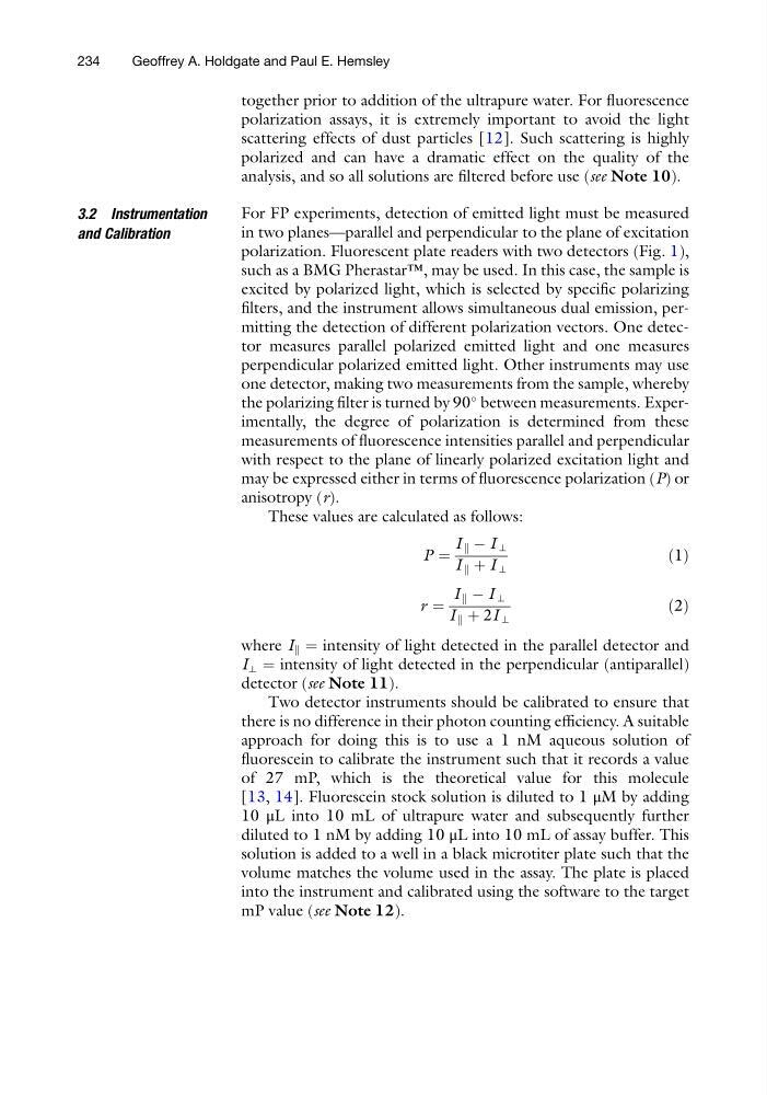
These values are calculated as follows:
P ¼
I k À I ⊥
I k þ I ⊥
ð1Þ
r ¼
I k À I ⊥
I k þ 2I ⊥
ð2Þ
where I k ¼ intensity of light detected in the parallel detector and
I ⊥ ¼ intensity of light detected in the perpendicular (antiparallel)
detector (see Note 11).
Two detector instruments should be calibrated to ensure that
there is no difference in their photon counting efficiency. A suitable
approach for doing this is to use a 1 nM aqueous solution of
fluorescein to calibrate the instrument such that it records a value
of 27 mP, which is the theoretical value for this molecule
[13, 14]. Fluorescein stock solution is diluted to 1 μM by adding
10 μL into 10 mL of ultrapure water and subsequently further
diluted to 1 nM by adding 10 μL into 10 mL of assay buffer. This
solution is added to a well in a black microtiter plate such that the
volume matches the volume used in the assay. The plate is placed
into the instrument and calibrated using the software to the target
mP value (see Note 12).
234
Geoffrey A. Holdgate and Paul E. Hemsley
polarization assays, it is extremely important to avoid the light
scattering effects of dust particles [12]. Such scattering is highly
polarized and can have a dramatic effect on the quality of the
analysis, and so all solutions are filtered before use (see Note 10).
3.2 Instrumentation
and Calibration
For FP experiments, detection of emitted light must be measured
in two planes—parallel and perpendicular to the plane of excitation
polarization. Fluorescent plate readers with two detectors (Fig. 1),
such as a BMG Pherastar™, may be used. In this case, the sample is
excited by polarized light, which is selected by specific polarizing
filters, and the instrument allows simultaneous dual emission, permitting the detection of different polarization vectors. One detector measures parallel polarized emitted light and one measures
perpendicular polarized emitted light. Other instruments may use
one detector, making two measurements from the sample, whereby
the polarizing filter is turned by 90
between measurements. Experimentally, the degree of polarization is determined from these
measurements of fluorescence intensities parallel and perpendicular
with respect to the plane of linearly polarized excitation light and
may be expressed either in terms of fluorescence polarization (P) or
anisotropy (r).
These values are calculated as follows:
P ¼
I k À I ⊥
I k þ I ⊥
ð1Þ
r ¼
I k À I ⊥
I k þ 2I ⊥
ð2Þ
where I k ¼ intensity of light detected in the parallel detector and
I ⊥ ¼ intensity of light detected in the perpendicular (antiparallel)
detector (see Note 11).
Two detector instruments should be calibrated to ensure that
there is no difference in their photon counting efficiency. A suitable
approach for doing this is to use a 1 nM aqueous solution of
fluorescein to calibrate the instrument such that it records a value
of 27 mP, which is the theoretical value for this molecule
[13, 14]. Fluorescein stock solution is diluted to 1 μM by adding
10 μL into 10 mL of ultrapure water and subsequently further
diluted to 1 nM by adding 10 μL into 10 mL of assay buffer. This
solution is added to a well in a black microtiter plate such that the
volume matches the volume used in the assay. The plate is placed
into the instrument and calibrated using the software to the target
mP value (see Note 12).
234
Geoffrey A. Holdgate and Paul E. Hemsley