

of 120 μg/mL and 30 μL anti-rabbit IgG (Fc specific) AlphaLISA Acceptor beads to give a concentration of 30 μg/mL.
3. Under subdued light, add 6 μL/well of the combined antibody
solution to the cell lysate in the AlphaScreen compatible
384-well plate, e.g., ProxiPlate-384.
4. Apply a plate seal.
5. Ensure mixing of antibodies with cell lysate by performing a
brief centrifugation at 300 Â g for 10 s.
6. Under subdued light, incubate plates for 4–16 h at room
temperature.
7. Analyze AlphaScreen signal using a compatible plate reader,
e.g., EnVision. Excite at 680 nm for 180 ms and measure
emission at 570 nm for 550 ms.
3.6 Screening
of Compounds
at a Single Optimized
Temperature by
CETSA HT
Volumes are provided for execution of one 384-well plate for
compound screening but can be scaled as appropriate. The optimal
target temperature for the heat shock will be determined from the
thermal melt analysis performed in Subheading 3.4. The optimal
temperature is the lowest available temperature, which gives a
suitable assay window between low AlphaScreen signal in cells
treated with DMSO control and high AlphaScreen signal in cells
treated with the tool compound. This is typically where 80–90% of
AlphaScreen signal has been lost in the DMSO control melt curve
(Fig. 1b).
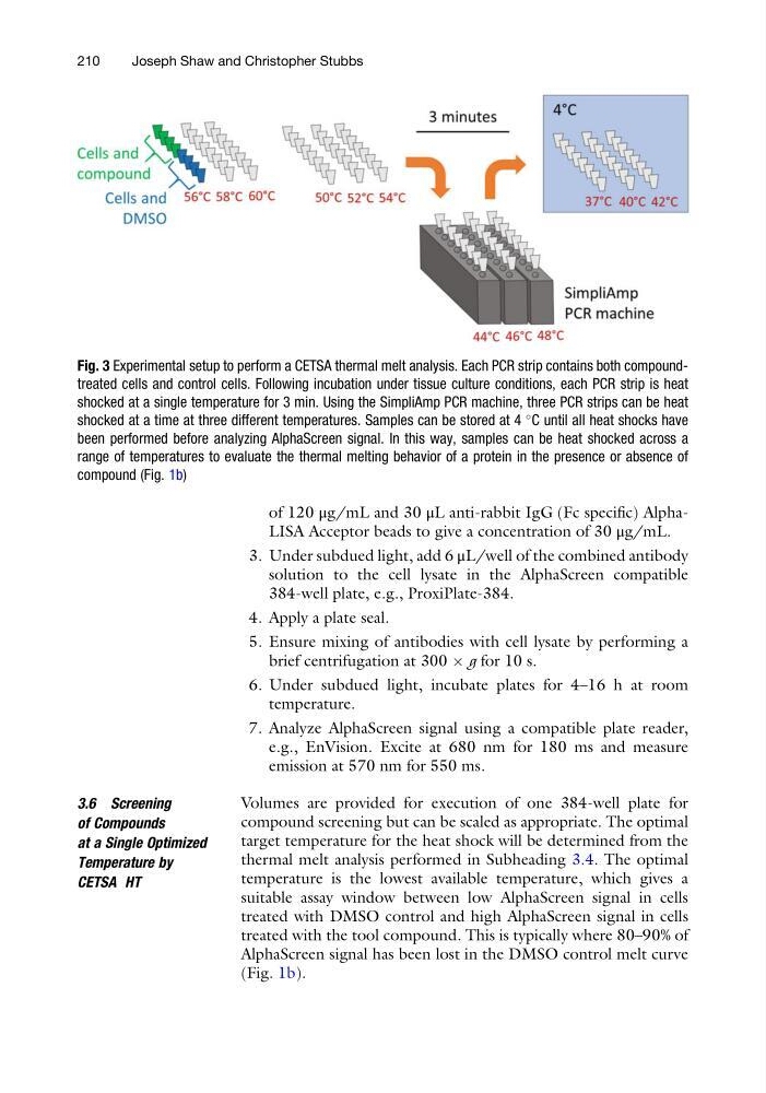
Fig. 3 Experimental setup to perform a CETSA thermal melt analysis. Each PCR strip contains both compoundtreated cells and control cells. Following incubation under tissue culture conditions, each PCR strip is heat
shocked at a single temperature for 3 min. Using the SimpliAmp PCR machine, three PCR strips can be heat
shocked at a time at three different temperatures. Samples can be stored at 4
C until all heat shocks have
been performed before analyzing AlphaScreen signal. In this way, samples can be heat shocked across a
range of temperatures to evaluate the thermal melting behavior of a protein in the presence or absence of
compound (Fig. 1b)
210
Joseph Shaw and Christopher Stubbs
3. Under subdued light, add 6 μL/well of the combined antibody
solution to the cell lysate in the AlphaScreen compatible
384-well plate, e.g., ProxiPlate-384.
4. Apply a plate seal.
5. Ensure mixing of antibodies with cell lysate by performing a
brief centrifugation at 300 Â g for 10 s.
6. Under subdued light, incubate plates for 4–16 h at room
temperature.
7. Analyze AlphaScreen signal using a compatible plate reader,
e.g., EnVision. Excite at 680 nm for 180 ms and measure
emission at 570 nm for 550 ms.
3.6 Screening
of Compounds
at a Single Optimized
Temperature by
CETSA HT
Volumes are provided for execution of one 384-well plate for
compound screening but can be scaled as appropriate. The optimal
target temperature for the heat shock will be determined from the
thermal melt analysis performed in Subheading 3.4. The optimal
temperature is the lowest available temperature, which gives a
suitable assay window between low AlphaScreen signal in cells
treated with DMSO control and high AlphaScreen signal in cells
treated with the tool compound. This is typically where 80–90% of
AlphaScreen signal has been lost in the DMSO control melt curve
(Fig. 1b).
Fig. 3 Experimental setup to perform a CETSA thermal melt analysis. Each PCR strip contains both compoundtreated cells and control cells. Following incubation under tissue culture conditions, each PCR strip is heat
shocked at a single temperature for 3 min. Using the SimpliAmp PCR machine, three PCR strips can be heat
shocked at a time at three different temperatures. Samples can be stored at 4
C until all heat shocks have
been performed before analyzing AlphaScreen signal. In this way, samples can be heat shocked across a
range of temperatures to evaluate the thermal melting behavior of a protein in the presence or absence of
compound (Fig. 1b)
210
Joseph Shaw and Christopher Stubbs