

4. Incubate at room temperature 5 min. The DNA precipitates on
the beads.
5. Place the PCR plate on a magnetic stand and wait for the
solution to become clear. At this point, the magnetic beads
form a small pellet on the side of the well facing the magnet.
6. Carefully aspirate the supernatant from each well.
7. Wash the beads by adding 200 μl 70% ethanol to each well and
incubate for 1 min.
8. Discard the supernatant and leave the plate to air-dry until the
beads are dry. It usually takes ~15 min until the bead pellets
start to crack.
9. Remove the PCR plate from the magnetic stand and elute the
amplicons using 12.5 μl buffer EB.
10. Mix by carefully pipetting up and down 15 times or until the
pellet is fully dissolved.
11. Incubate for 5 min at room temperature.
12. Place the PCR plate on the magnetic stand and wait for
the solution to become clear.
13. Combine 10 μl of each amplicon into a single 1.5 ml tube (see
Note 7).
14. Concentrate the pooled PCR products using a QIAgen PCR
cleanup column according to the manual. Elute in 50 μl TE
buffer.
15. Purify the PCR product by agarose gel electrophoresis and.
Prepare a 2% agarose gel with 60 μl wells and run the gel at
80 mA for 40 min.
16. Excise the band of expected size, taking care not to include
shorter fragments.
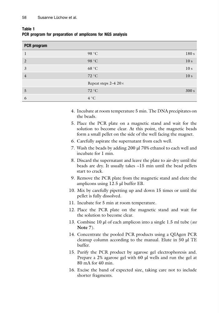
Table 1
PCR program for preparation of amplicons for NGS analysis
PCR program
1
9 8
C
180 s
2
9 8
C
1 0 s
3
6 8
C
1 0 s
4
7 2
C
1 0 s
Repeat steps 2–4 20Â
5
7 2
C
300 s
6
4
C
58
Susanne Lu ¨ chow et al.
the beads.
5. Place the PCR plate on a magnetic stand and wait for the
solution to become clear. At this point, the magnetic beads
form a small pellet on the side of the well facing the magnet.
6. Carefully aspirate the supernatant from each well.
7. Wash the beads by adding 200 μl 70% ethanol to each well and
incubate for 1 min.
8. Discard the supernatant and leave the plate to air-dry until the
beads are dry. It usually takes ~15 min until the bead pellets
start to crack.
9. Remove the PCR plate from the magnetic stand and elute the
amplicons using 12.5 μl buffer EB.
10. Mix by carefully pipetting up and down 15 times or until the
pellet is fully dissolved.
11. Incubate for 5 min at room temperature.
12. Place the PCR plate on the magnetic stand and wait for
the solution to become clear.
13. Combine 10 μl of each amplicon into a single 1.5 ml tube (see
Note 7).
14. Concentrate the pooled PCR products using a QIAgen PCR
cleanup column according to the manual. Elute in 50 μl TE
buffer.
15. Purify the PCR product by agarose gel electrophoresis and.
Prepare a 2% agarose gel with 60 μl wells and run the gel at
80 mA for 40 min.
16. Excise the band of expected size, taking care not to include
shorter fragments.
Table 1
PCR program for preparation of amplicons for NGS analysis
PCR program
1
9 8
C
180 s
2
9 8
C
1 0 s
3
6 8
C
1 0 s
4
7 2
C
1 0 s
Repeat steps 2–4 20Â
5
7 2
C
300 s
6
4
C
58
Susanne Lu ¨ chow et al.