

C-terminal carboxylate group from the protein partner interacts
with the backbone of a conserved loop in the PDZ domain through
a canonically conserved hydrogen bond network [2].
More precisely, PDZ domains typically require a hydrophobic
residue at the C-terminal position of their protein partner, and this
residue fits into a small hydrophobic pocket near the carboxylate
binding loop. Typical hydrophobic residues are valine, leucine and
isoleucine. By convention, P0 refers to the C-terminal residue of
the peptide, and P-n refers to the nth amino acid of the peptide
starting from its C-terminal end. PDZ domains have been classified
according to their specificity for PDZ ligands. Consensus sequences
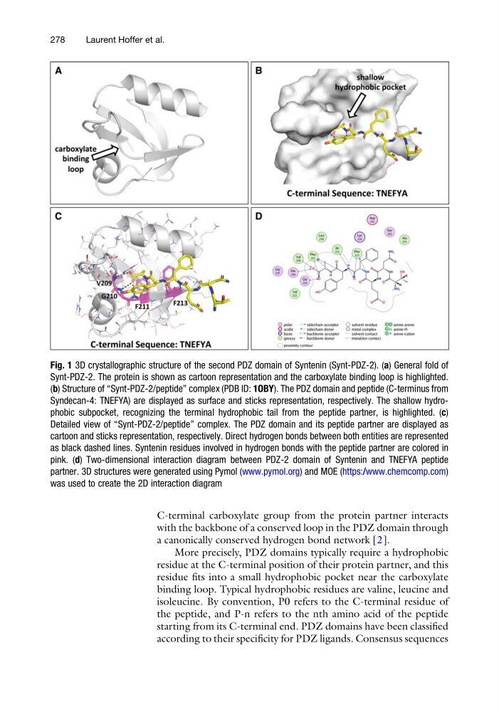
Fig. 1 3D crystallographic structure of the second PDZ domain of Syntenin (Synt-PDZ-2). (a) General fold of
Synt-PDZ-2. The protein is shown as cartoon representation and the carboxylate binding loop is highlighted.
(b) Structure of “Synt-PDZ-2/peptide” complex (PDB ID: 1OBY). The PDZ domain and peptide (C-terminus from
Syndecan-4: TNEFYA) are displayed as surface and sticks representation, respectively. The shallow hydrophobic subpocket, recognizing the terminal hydrophobic tail from the peptide partner, is highlighted. (c)
Detailed view of “Synt-PDZ-2/peptide” complex. The PDZ domain and its peptide partner are displayed as
cartoon and sticks representation, respectively. Direct hydrogen bonds between both entities are represented
as black dashed lines. Syntenin residues involved in hydrogen bonds with the peptide partner are colored in
pink. (d) Two-dimensional interaction diagram between PDZ-2 domain of Syntenin and TNEFYA peptide
partner. 3D structures were generated using Pymol (www.pymol.org) and MOE (https:/ /www.chemcomp.com)
was used to create the 2D interaction diagram
278
Laurent Hoffer et al.
with the backbone of a conserved loop in the PDZ domain through
a canonically conserved hydrogen bond network [2].
More precisely, PDZ domains typically require a hydrophobic
residue at the C-terminal position of their protein partner, and this
residue fits into a small hydrophobic pocket near the carboxylate
binding loop. Typical hydrophobic residues are valine, leucine and
isoleucine. By convention, P0 refers to the C-terminal residue of
the peptide, and P-n refers to the nth amino acid of the peptide
starting from its C-terminal end. PDZ domains have been classified
according to their specificity for PDZ ligands. Consensus sequences
Fig. 1 3D crystallographic structure of the second PDZ domain of Syntenin (Synt-PDZ-2). (a) General fold of
Synt-PDZ-2. The protein is shown as cartoon representation and the carboxylate binding loop is highlighted.
(b) Structure of “Synt-PDZ-2/peptide” complex (PDB ID: 1OBY). The PDZ domain and peptide (C-terminus from
Syndecan-4: TNEFYA) are displayed as surface and sticks representation, respectively. The shallow hydrophobic subpocket, recognizing the terminal hydrophobic tail from the peptide partner, is highlighted. (c)
Detailed view of “Synt-PDZ-2/peptide” complex. The PDZ domain and its peptide partner are displayed as
cartoon and sticks representation, respectively. Direct hydrogen bonds between both entities are represented
as black dashed lines. Syntenin residues involved in hydrogen bonds with the peptide partner are colored in
pink. (d) Two-dimensional interaction diagram between PDZ-2 domain of Syntenin and TNEFYA peptide
partner. 3D structures were generated using Pymol (www.pymol.org) and MOE (https:/ /www.chemcomp.com)
was used to create the 2D interaction diagram
278
Laurent Hoffer et al.