

from genera Alphacoronavirus and Betacoronavirus. However, this
was not the case in CoVs evolved from birds with the exception of E
protein from bottlenose dolphin CoV HKU22. Interestingly,
SARS-CoV, MERS-CoV, and SARS-CoV-2, the three human
CoVs that are highly pathogenic, have three proteins including a
PBM in their carboxy-terminus: proteins E, 3a and 7b in SARSCoV, proteins E, 5 and M in MERS-CoV and proteins E, 3a and
8 in SARS-CoV-2. In addition, MERS-CoV is the only CoV in
which a PBM has been described in M protein, a structural CoV
protein essential for viral assembly [73]. Remarkably, MERS-CoV
proteins E and 5 are homologs of SARS-CoV and SARS-CoV2 proteins E and 3a, respectively, suggesting that they may play a
similar role in MERS-CoV. Furthermore, SARS-CoV and MERSCoV, as most Betacoronaviruses, were originated in bats and then
transmitted to humans through intermediate hosts: civet cats in the
case of SARS-CoV, and camels in the case of MERS-CoV. GenBank
data from genomes of hundreds of SARS-CoVs and MERS-CoVs
variants isolated from bats, civets, camels and humans were analyzed showing that the PBMs from SARS-CoV E and 3a proteins
were mostly conserved in bats, civets and humans similarly to the
PBMs of MERS-CoV proteins E, 5 that were also mostly conserved
in bats, camels and humans (Table 3, adapted from [67]).
Interestingly, the mutations that affected the PBM core
sequences introduced a different PBM, likely functional in the
corresponding animal context. This phylogenetic conservation
reinforces the relevance of viral PBMs in CoVs opening the possibility that they could be involved in CoV adaptation to the host.
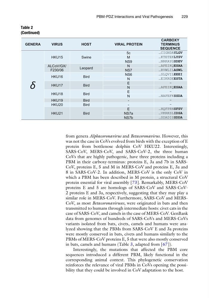
Table 2
(Continued)
GENERA
VIRUS
HOST
VIRAL PROTEIN
CARBOXY
TERMINUS
SEQUENCE
δ
HKU15
Swine
5c
…CIGNDAYLGV
M
…KTETEKLYSV
NS9
…NRKAYGSDEV
ALCoV/GX/
F230/06
Leopard
N
…AFEIKQESAA
NS7
…RVWLILASWL
HKU16
Bird
NS6
…SLQVILEEEI
N
…EIKRDEESTA
HKU17
Bird
E
-
N
…AFEIKQESAA
HKU18
Bird
E
-
N
…NAFEFKSSDA
HKU19
Bird
-
-
HKU20
Bird
-
-
HKU21
Bird
E
…HQFPRNSFSV
NS7a
…VKRKSLIDSA
NS7b
…SDADISSDDA
PBM-PDZ Interactions and Viral Pathogenesis
229
was not the case in CoVs evolved from birds with the exception of E
protein from bottlenose dolphin CoV HKU22. Interestingly,
SARS-CoV, MERS-CoV, and SARS-CoV-2, the three human
CoVs that are highly pathogenic, have three proteins including a
PBM in their carboxy-terminus: proteins E, 3a and 7b in SARSCoV, proteins E, 5 and M in MERS-CoV and proteins E, 3a and
8 in SARS-CoV-2. In addition, MERS-CoV is the only CoV in
which a PBM has been described in M protein, a structural CoV
protein essential for viral assembly [73]. Remarkably, MERS-CoV
proteins E and 5 are homologs of SARS-CoV and SARS-CoV2 proteins E and 3a, respectively, suggesting that they may play a
similar role in MERS-CoV. Furthermore, SARS-CoV and MERSCoV, as most Betacoronaviruses, were originated in bats and then
transmitted to humans through intermediate hosts: civet cats in the
case of SARS-CoV, and camels in the case of MERS-CoV. GenBank
data from genomes of hundreds of SARS-CoVs and MERS-CoVs
variants isolated from bats, civets, camels and humans were analyzed showing that the PBMs from SARS-CoV E and 3a proteins
were mostly conserved in bats, civets and humans similarly to the
PBMs of MERS-CoV proteins E, 5 that were also mostly conserved
in bats, camels and humans (Table 3, adapted from [67]).
Interestingly, the mutations that affected the PBM core
sequences introduced a different PBM, likely functional in the
corresponding animal context. This phylogenetic conservation
reinforces the relevance of viral PBMs in CoVs opening the possibility that they could be involved in CoV adaptation to the host.
Table 2
(Continued)
GENERA
VIRUS
HOST
VIRAL PROTEIN
CARBOXY
TERMINUS
SEQUENCE
δ
HKU15
Swine
5c
…CIGNDAYLGV
M
…KTETEKLYSV
NS9
…NRKAYGSDEV
ALCoV/GX/
F230/06
Leopard
N
…AFEIKQESAA
NS7
…RVWLILASWL
HKU16
Bird
NS6
…SLQVILEEEI
N
…EIKRDEESTA
HKU17
Bird
E
-
N
…AFEIKQESAA
HKU18
Bird
E
-
N
…NAFEFKSSDA
HKU19
Bird
-
-
HKU20
Bird
-
-
HKU21
Bird
E
…HQFPRNSFSV
NS7a
…VKRKSLIDSA
NS7b
…SDADISSDDA
PBM-PDZ Interactions and Viral Pathogenesis
229