

5. Incubate the transfection mixture on room temperature for at
least 10 min.
6. Add 8 μl of transfection mixture to each well and incubate the
plate at 37
C, 5% CO 2 for 4–6 h.
7. To monitor signal transduction, remove carefully the medium
from each well and add 100 μl of serum-free DMEM for
washing out FBS (see Note 13).
8. Remove the washing medium and add 100 μl of CO 2 independent medium (supplemented with L-glutamine and
antibiotics).
9. Incubate the plate at 37
C with 5% CO 2 for further 18–20 h
(see Note 14).
10. Set the plate reader to 37
C (optional).
11. Prepare the Nano-Glo Live Cell reagent as follows: Melt the
Nano-Glo LCS Dilution buffer and the Nano-Glo Live Cell
Substrate on ice. For 6 wells, add 8.75 μl Nano-Glo Live Cell
Substrate to 166.25 μl Nano-Glo LCS Dilution buffer, mix it
gently by pipetting (see Note 15).
12. Add 25 μl of Nano-Glo Live Cell reagent to each well, then
start measuring the luminescence with 1 min interval. Record
the baseline until luminescence begins to decrease (see Note
16).
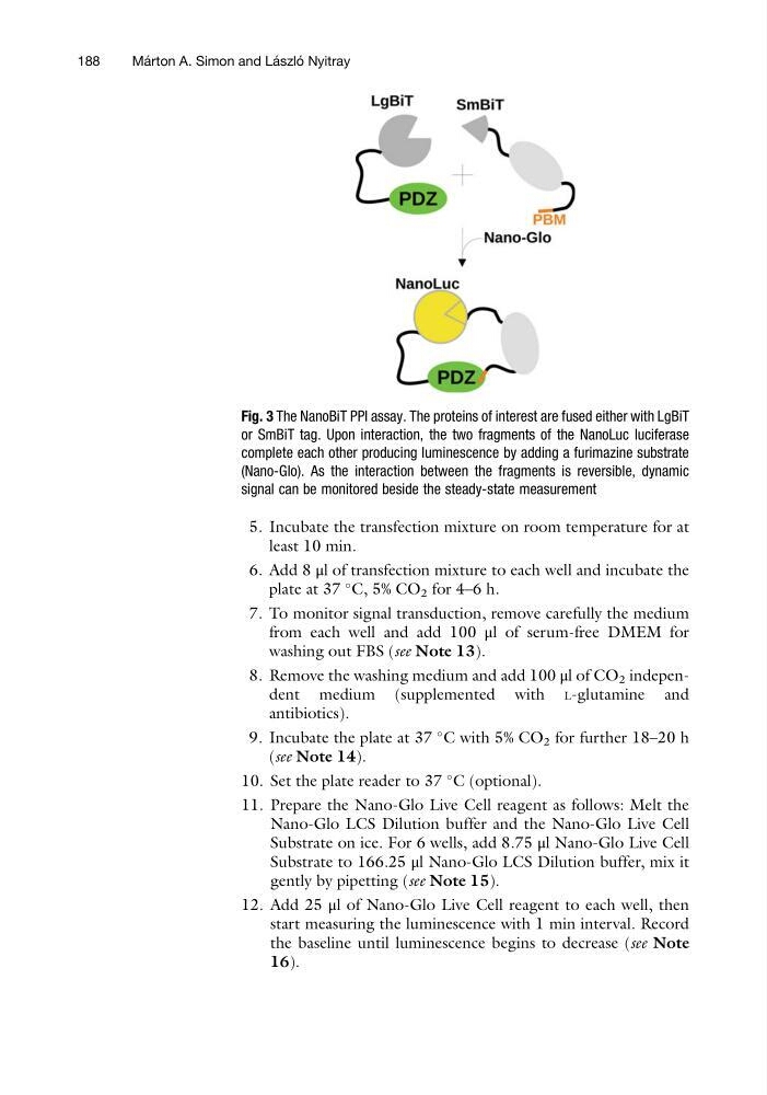
Fig. 3 The NanoBiT PPI assay. The proteins of interest are fused either with LgBiT
or SmBiT tag. Upon interaction, the two fragments of the NanoLuc luciferase
complete each other producing luminescence by adding a furimazine substrate
(Nano-Glo). As the interaction between the fragments is reversible, dynamic
signal can be monitored beside the steady-state measurement
188
Ma ´ rton A. Simon and La ´ szlo ´ Nyitray
least 10 min.
6. Add 8 μl of transfection mixture to each well and incubate the
plate at 37
C, 5% CO 2 for 4–6 h.
7. To monitor signal transduction, remove carefully the medium
from each well and add 100 μl of serum-free DMEM for
washing out FBS (see Note 13).
8. Remove the washing medium and add 100 μl of CO 2 independent medium (supplemented with L-glutamine and
antibiotics).
9. Incubate the plate at 37
C with 5% CO 2 for further 18–20 h
(see Note 14).
10. Set the plate reader to 37
C (optional).
11. Prepare the Nano-Glo Live Cell reagent as follows: Melt the
Nano-Glo LCS Dilution buffer and the Nano-Glo Live Cell
Substrate on ice. For 6 wells, add 8.75 μl Nano-Glo Live Cell
Substrate to 166.25 μl Nano-Glo LCS Dilution buffer, mix it
gently by pipetting (see Note 15).
12. Add 25 μl of Nano-Glo Live Cell reagent to each well, then
start measuring the luminescence with 1 min interval. Record
the baseline until luminescence begins to decrease (see Note
16).
Fig. 3 The NanoBiT PPI assay. The proteins of interest are fused either with LgBiT
or SmBiT tag. Upon interaction, the two fragments of the NanoLuc luciferase
complete each other producing luminescence by adding a furimazine substrate
(Nano-Glo). As the interaction between the fragments is reversible, dynamic
signal can be monitored beside the steady-state measurement
188
Ma ´ rton A. Simon and La ´ szlo ´ Nyitray