

2.4.3 NMR
1. 600 MHz (or higher fields) NMR spectrometer equipped with
triple resonance
1
H{
13
C/
15 N} PFG probe (for heteronuclear
NMR detection).
2. 4 mm NMR tube (Shigemi INC).
3. 100 μM PDZ sample (250 μl).
4. 100% D 2 O to add to the PDZ sample at 1–5% final
concentration.
3 Methods
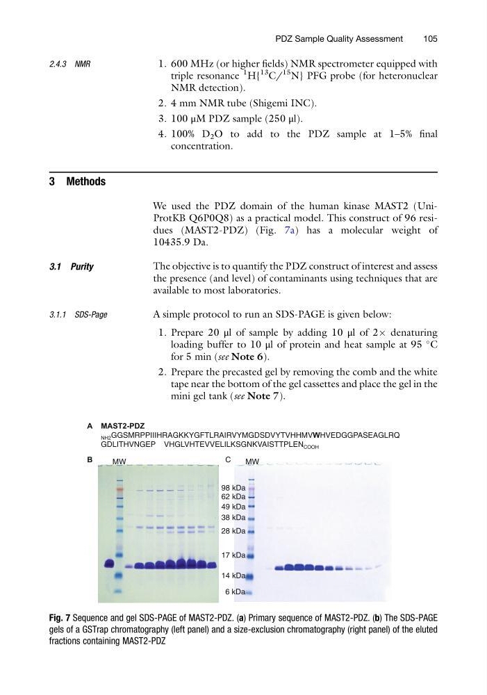
We used the PDZ domain of the human kinase MAST2 (UniProtKB Q6P0Q8) as a practical model. This construct of 96 residues (MAST2-PDZ) (Fig. 7a) has a molecular weight of
10435.9 Da.
3.1 Purity
The objective is to quantify the PDZ construct of interest and assess
the presence (and level) of contaminants using techniques that are
available to most laboratories.
3.1.1 SDS-Page
A simple protocol to run an SDS-PAGE is given below:
1. Prepare 20 μl of sample by adding 10 μl of 2Â denaturing
loading buffer to 10 μl of protein and heat sample at 95
C
for 5 min (see Note 6).
2. Prepare the precasted gel by removing the comb and the white
tape near the bottom of the gel cassettes and place the gel in the
mini gel tank (see Note 7).
MAST2-PDZ
NH2 GGSMRPPIIIHRAGKKYGFTLRAIRVYMGDSDVYTVHHMVWHVEDGGPASEAGLRQ
GDLITHVNGEP VHGLVHTEVVELILKSGNKVAISTTPLEN COOH
6 kDa
14 kDa
17 kDa
28 kDa
38 kDa
49 kDa
62 kDa
98 kDa
MW
MW
B
C
A
Fig. 7 Sequence and gel SDS-PAGE of MAST2-PDZ. (a) Primary sequence of MAST2-PDZ. (b) The SDS-PAGE
gels of a GSTrap chromatography (left panel) and a size-exclusion chromatography (right panel) of the eluted
fractions containing MAST2-PDZ
PDZ Sample Quality Assessment
105
1. 600 MHz (or higher fields) NMR spectrometer equipped with
triple resonance
1
H{
13
C/
15 N} PFG probe (for heteronuclear
NMR detection).
2. 4 mm NMR tube (Shigemi INC).
3. 100 μM PDZ sample (250 μl).
4. 100% D 2 O to add to the PDZ sample at 1–5% final
concentration.
3 Methods
We used the PDZ domain of the human kinase MAST2 (UniProtKB Q6P0Q8) as a practical model. This construct of 96 residues (MAST2-PDZ) (Fig. 7a) has a molecular weight of
10435.9 Da.
3.1 Purity
The objective is to quantify the PDZ construct of interest and assess
the presence (and level) of contaminants using techniques that are
available to most laboratories.
3.1.1 SDS-Page
A simple protocol to run an SDS-PAGE is given below:
1. Prepare 20 μl of sample by adding 10 μl of 2Â denaturing
loading buffer to 10 μl of protein and heat sample at 95
C
for 5 min (see Note 6).
2. Prepare the precasted gel by removing the comb and the white
tape near the bottom of the gel cassettes and place the gel in the
mini gel tank (see Note 7).
MAST2-PDZ
NH2 GGSMRPPIIIHRAGKKYGFTLRAIRVYMGDSDVYTVHHMVWHVEDGGPASEAGLRQ
GDLITHVNGEP VHGLVHTEVVELILKSGNKVAISTTPLEN COOH
6 kDa
14 kDa
17 kDa
28 kDa
38 kDa
49 kDa
62 kDa
98 kDa
MW
MW
B
C
A
Fig. 7 Sequence and gel SDS-PAGE of MAST2-PDZ. (a) Primary sequence of MAST2-PDZ. (b) The SDS-PAGE
gels of a GSTrap chromatography (left panel) and a size-exclusion chromatography (right panel) of the eluted
fractions containing MAST2-PDZ
PDZ Sample Quality Assessment
105