

7. Once cells are returned to the BSL-3, they are ready to be
prepped for Alamar Blue viability assays (see Note 28).
8. Fill a 25 Â 150 mm glass tube with 10 mL of 7H9
medium + OADC + 0.05% tween and a small magnetic stir bar.
9. Using a sterile, disposable inoculation loop, add four loops of
inactivated M. tuberculosis cells to the 25 Â 150 mm glass tube
containing the sterile medium and small magnetic stir bar.
Label as “Test sample” (you may have multiple test samples).
10. Transfer 1 mL of an actively growing M. tuberculosis culture
into another 25 Â 150 mm glass tube containing the sterile
medium and small magnetic stir bar (see Note 29), label as
“positive control.”
11. Obtain another 25 Â 150 mm glass tube containing the sterile
medium and small magnetic stir bar and label as “blank” (see
Note 30).
12. Place tubes on a magnetic stir plate at 37
C for 72 h.
13. Remove samples and read OD 600 .
14. Dilute the positive control and test sample(s) to OD 600 ¼ 1.0
using sterile medium.
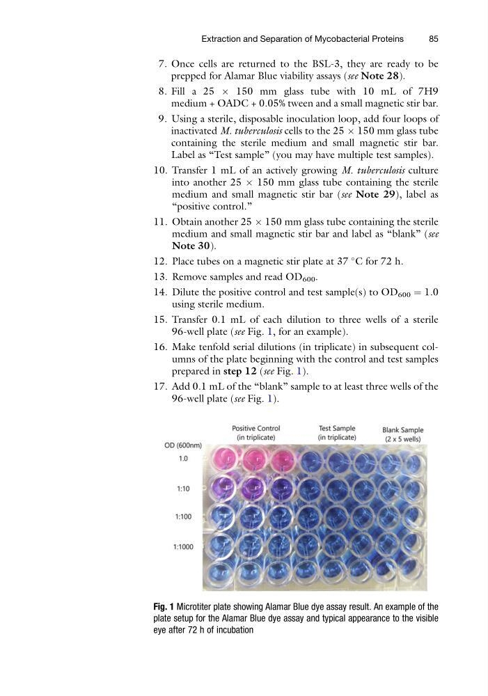
15. Transfer 0.1 mL of each dilution to three wells of a sterile
96-well plate (see Fig. 1, for an example).
16. Make tenfold serial dilutions (in triplicate) in subsequent columns of the plate beginning with the control and test samples
prepared in step 12 (see Fig. 1).
17. Add 0.1 mL of the “blank” sample to at least three wells of the
96-well plate (see Fig. 1).
Fig. 1 Microtiter plate showing Alamar Blue dye assay result. An example of the
plate setup for the Alamar Blue dye assay and typical appearance to the visible
eye after 72 h of incubation
Extraction and Separation of Mycobacterial Proteins
85
prepped for Alamar Blue viability assays (see Note 28).
8. Fill a 25 Â 150 mm glass tube with 10 mL of 7H9
medium + OADC + 0.05% tween and a small magnetic stir bar.
9. Using a sterile, disposable inoculation loop, add four loops of
inactivated M. tuberculosis cells to the 25 Â 150 mm glass tube
containing the sterile medium and small magnetic stir bar.
Label as “Test sample” (you may have multiple test samples).
10. Transfer 1 mL of an actively growing M. tuberculosis culture
into another 25 Â 150 mm glass tube containing the sterile
medium and small magnetic stir bar (see Note 29), label as
“positive control.”
11. Obtain another 25 Â 150 mm glass tube containing the sterile
medium and small magnetic stir bar and label as “blank” (see
Note 30).
12. Place tubes on a magnetic stir plate at 37
C for 72 h.
13. Remove samples and read OD 600 .
14. Dilute the positive control and test sample(s) to OD 600 ¼ 1.0
using sterile medium.
15. Transfer 0.1 mL of each dilution to three wells of a sterile
96-well plate (see Fig. 1, for an example).
16. Make tenfold serial dilutions (in triplicate) in subsequent columns of the plate beginning with the control and test samples
prepared in step 12 (see Fig. 1).
17. Add 0.1 mL of the “blank” sample to at least three wells of the
96-well plate (see Fig. 1).
Fig. 1 Microtiter plate showing Alamar Blue dye assay result. An example of the
plate setup for the Alamar Blue dye assay and typical appearance to the visible
eye after 72 h of incubation
Extraction and Separation of Mycobacterial Proteins
85