

extra-capillary space (ECS) formed between the fiber and the inner
surface of the cylinder. Cells may be cultured in isolation within this
secondary ECS compartment because the cells are too large to pass
through the pores of the fiber walls into the primary flow path
inside the fiber. Thus, the researcher has dynamic control of the
environmental growth conditions as well as the supply/removal of
nutrients, drugs, and other factors.
The use of hollow fiber bioreactor technology to study the
bactericidal effects of antibiotics was first reported in 1981 [1],
and 3 years later it was applied to model the pharmacodynamics
(PD) of fluoroquinolone interaction with Mycobacterium tuberculosis in order to optimize treatment while minimizing resistance
development [2]. The strength of the HF model of tuberculosis
lies in its ability to accurately simulate an animal or human PK/PD
relationship with one or more drugs that may have different halflives. Furthermore, it is a biosafe system since the pathogenic
bacteria are sequestered in an isolated compartment. Samples can
be taken from the bacteria-free CR to monitor circulating drug
levels or from the ECS to follow bacterial viability—even to the
point of complete sterility. In either case, taking small sample
volumes has negligible impact on growth of the culture itself.
As an in vitro testing system, the HF model also has major
limitations. Foremost is the absence of host-derived factors, including TB-specific elements of the immune response, as well as the
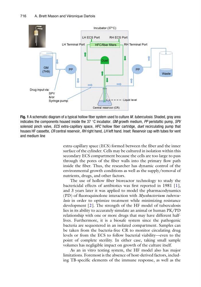
Fig. 1 A schematic diagram of a typical hollow fiber system used to culture M. tuberculosis. Shaded, gray area
indicates the components housed inside the 37
C incubator. GM growth medium, PP peristaltic pump, SPV
solenoid pinch valve, ECS extra-capillary space, HFC hollow fiber cartridge, duet recirculating pump that
houses HF cassette, CR central reservoir, RH right hand, LH left hand. Inset: Reservoir cap with tubes for vent
and medium line
716
A. Brett Mason and Ve ´ ronique Dartois