

4. Prepare assay plates by transferring compounds directly from
the source plate, using a high precision automatic liquid dispenser (see Note 9). The final amount of DMSO in the assay
plate should be <1% v/v for each well.
5. Store assay and source plates at À20
C, sealed with an adhesive
aluminum sealing film, until the day of infection.
6. Prepare a bacterial suspension at 2 Â 10
6 CFU/mL (see Subheading 3.1 and Note 18). A volume of 10 mL of bacterial
suspension per plate is required.
7. Prepare a cellular suspension as previously described (see Subheadings 3.2–3.4 and 3.7–3.10) to have the cell type-adapted
MOI (i.e. a cell suspension at 1 Â 10
6 cells/mL to obtain MOI
2 for RAW264.7 and THP-1 macrophage cell lines or 2 Â 10
6
cells/mL to obtain MOI 1 for human and murine primary
macrophages cells) (see Notes 18 and 20). A volume of
10 mL of cellular suspension per plate is required.
8. Mix one volume of cells with one volume of bacteria in a sterile
and ventilated bacterial culture Erlenmeyer of appropriate
volume.
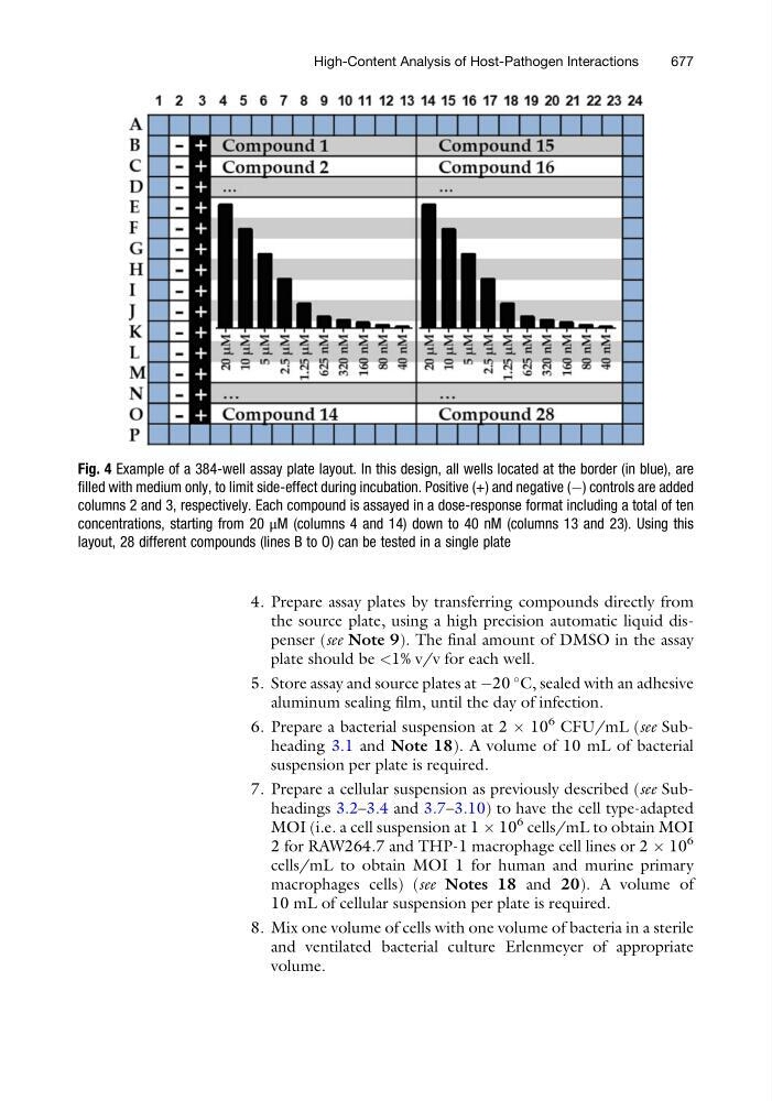
Fig. 4 Example of a 384-well assay plate layout. In this design, all wells located at the border (in blue), are
filled with medium only, to limit side-effect during incubation. Positive (+) and negative (À) controls are added
columns 2 and 3, respectively. Each compound is assayed in a dose-response format including a total of ten
concentrations, starting from 20 μM (columns 4 and 14) down to 40 nM (columns 13 and 23). Using this
layout, 28 different compounds (lines B to O) can be tested in a single plate
High-Content Analysis of Host-Pathogen Interactions
677