

high-resolution capacity that can now be obtained with modern
devices, not only can quantification of bacterial survival be
recorded, but phenotypic changes of either the bacteria or the
host cell can also be observed simultaneously. The methodology
presented here was developed to quantify intracellular
M. tuberculosis by detecting enhanced-GFP (eGFP)-expressing bacteria inside fluorescently labeled phagocytic or non-phagocytic
cells.
The first part of this chapter explains the preparation and use of
the reagents and the equipment required for the different assays. In
the second part, the cultivation methodology of selected cell lines
and primary cells are provided, which we use successfully for the
investigation of M. tuberculosis infection in our laboratory. The
robustness and rapid growth of the murine macrophage cell line
RAW264.7 make this cell model a good candidate for large-scale
screens. However, readers should consider differences in cell surface receptor expression and the different composition of the phagosomal proteome between RAW264.7 and primary macrophages
[13], when choosing their experimental model. More specific assays
have been efficaciously adapted in our laboratory to other cell lines,
such as the human monocyte cell line THP-1, the A549 human
type II pneumocyte-like cell line, murine bone marrow-derived
macrophages or dendritic cells, and human peripheral bloodM. tuberculosis
Lipid bodies
accumulation
Metal
intoxication
Phagosome
acidification
Vacuole
Cu +
Zn +
Block of phagosome
acidification
NO/ROS efflux
Replication
Vacuole
rupture
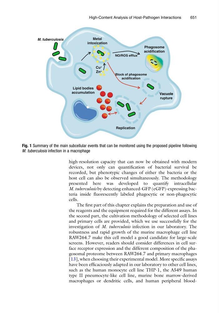
Fig. 1 Summary of the main subcellular events that can be monitored using the proposed pipeline following
M. tuberculosis infection in a macrophage
High-Content Analysis of Host-Pathogen Interactions
651