

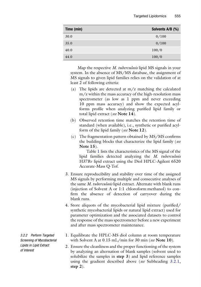
Time (min)
Solvents A/B (%)
30.0
0/100
35.0
0/100
40.0
100/0
44.0
100/0
Map the respective M. tuberculosis lipid MS signals in your
system. In the absence of MS/MS database, the assignment of
MS signals to given lipid families relies on the validation of at
least 2 of following criteria:
(a) The lipids are detected at m/z matching the calculated
m/z within the mass accuracy of the high-resolution mass
spectrometer (as low as 1 ppm and never exceeding
10 ppm mass accuracy) and show the expected acylforms profile when analyzing purified lipid family or
total lipid extract (see Note 14).
(b) Observed retention time matches the retention time of
standard (when available), i.e., synthetic or purified acylform of the lipid family (see Note 12).
(c) The fragmentation pattern obtained by MS/MS confirms
the building blocks that characterize the lipid family (see
Note 15).
Table 1 lists the characteristics of the MS signal of the
lipid families detected analyzing the M. tuberculosis
H37Rv lipid extract using the Diol HPLC-Agilent 6520
Accurate-Mass Q-Tof.
3. Ensure reproducibility and stability over time of the assigned
MS signals by performing multiple and consecutive analyses of
the same M. tuberculosis lipid extract. Alternate with blank runs
(injection of Solvent A or 1:1 chloroform:methanol) to confirm the absence of detection of carryover during the
blank runs.
4. Store aliquots of the mycobacterial lipid mixture (purified/
synthetic mycobacterial lipids or natural lipid extract) used for
parameter optimization and the associated datasets to control
the response of the mass spectrometer before a new experiment
and after mass spectrometer maintenance.
3.2.2 Perform Targeted
Screening of Mycobacterial
Lipids in Lipid Extract
of Interest
1. Equilibrate the HPLC-MS diol column at room temperature
with Solvent A at 0.15 mL/min for 30 min (see Note 10).
2. Ensure the cleanliness and the proper functioning of the system
by analyzing an alternation of blank samples (solvent used to
solubilize the samples in step 3) and lipid reference samples
using the gradient described above (see Subheading 3.2.1,
step 2).
Targeted Lipidomics
555
Solvents A/B (%)
30.0
0/100
35.0
0/100
40.0
100/0
44.0
100/0
Map the respective M. tuberculosis lipid MS signals in your
system. In the absence of MS/MS database, the assignment of
MS signals to given lipid families relies on the validation of at
least 2 of following criteria:
(a) The lipids are detected at m/z matching the calculated
m/z within the mass accuracy of the high-resolution mass
spectrometer (as low as 1 ppm and never exceeding
10 ppm mass accuracy) and show the expected acylforms profile when analyzing purified lipid family or
total lipid extract (see Note 14).
(b) Observed retention time matches the retention time of
standard (when available), i.e., synthetic or purified acylform of the lipid family (see Note 12).
(c) The fragmentation pattern obtained by MS/MS confirms
the building blocks that characterize the lipid family (see
Note 15).
Table 1 lists the characteristics of the MS signal of the
lipid families detected analyzing the M. tuberculosis
H37Rv lipid extract using the Diol HPLC-Agilent 6520
Accurate-Mass Q-Tof.
3. Ensure reproducibility and stability over time of the assigned
MS signals by performing multiple and consecutive analyses of
the same M. tuberculosis lipid extract. Alternate with blank runs
(injection of Solvent A or 1:1 chloroform:methanol) to confirm the absence of detection of carryover during the
blank runs.
4. Store aliquots of the mycobacterial lipid mixture (purified/
synthetic mycobacterial lipids or natural lipid extract) used for
parameter optimization and the associated datasets to control
the response of the mass spectrometer before a new experiment
and after mass spectrometer maintenance.
3.2.2 Perform Targeted
Screening of Mycobacterial
Lipids in Lipid Extract
of Interest
1. Equilibrate the HPLC-MS diol column at room temperature
with Solvent A at 0.15 mL/min for 30 min (see Note 10).
2. Ensure the cleanliness and the proper functioning of the system
by analyzing an alternation of blank samples (solvent used to
solubilize the samples in step 3) and lipid reference samples
using the gradient described above (see Subheading 3.2.1,
step 2).
Targeted Lipidomics
555