

3. Add 5 mL of collagenase IV digestion solution (250 U/mL) to
each minced sample and transfer to a 15 mL conical tube.
4. Place the tubes in a zipper storage bag and seal to prevent
potential leakage, before incubating at 37
C on a nutator for
at least 45 min.
5. Pass digested samples through a 70 μm cell strainer placed on a
50 mL conical tube.
6. Centrifuge the sample at 1500 rpm (450 Â g) for 10 min.
7. Discard the supernatant without disturbing the pellet, and
resuspend the pellet in 5 mL ACK lysing buffer.
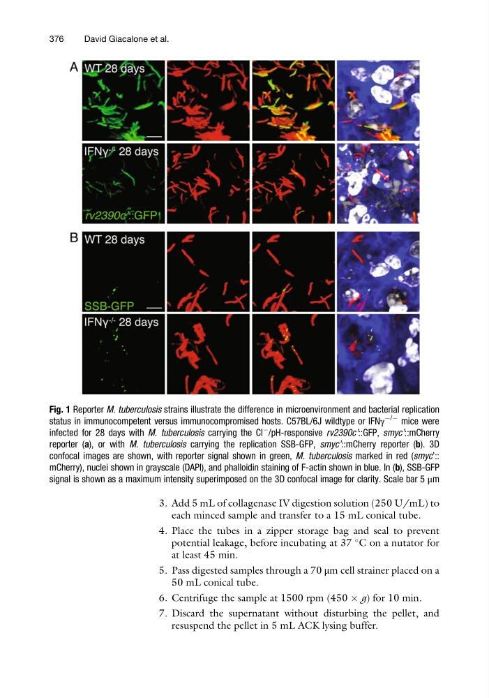
Fig. 1 Reporter M. tuberculosis strains illustrate the difference in microenvironment and bacterial replication
status in immunocompetent versus immunocompromised hosts. C57BL/6J wildtype or IFNγ
À/À
mice were
infected for 28 days with M. tuberculosis carrying the Cl
À /pH-responsive rv2390c’::GFP, smyc’::mCherry
reporter (a), or with M. tuberculosis carrying the replication SSB-GFP, smyc’::mCherry reporter (b). 3D
confocal images are shown, with reporter signal shown in green, M. tuberculosis marked in red (smyc’::
mCherry), nuclei shown in grayscale (DAPI), and phalloidin staining of F-actin shown in blue. In (b), SSB-GFP
signal is shown as a maximum intensity superimposed on the 3D confocal image for clarity. Scale bar 5 μm
376
David Giacalone et al.
each minced sample and transfer to a 15 mL conical tube.
4. Place the tubes in a zipper storage bag and seal to prevent
potential leakage, before incubating at 37
C on a nutator for
at least 45 min.
5. Pass digested samples through a 70 μm cell strainer placed on a
50 mL conical tube.
6. Centrifuge the sample at 1500 rpm (450 Â g) for 10 min.
7. Discard the supernatant without disturbing the pellet, and
resuspend the pellet in 5 mL ACK lysing buffer.
Fig. 1 Reporter M. tuberculosis strains illustrate the difference in microenvironment and bacterial replication
status in immunocompetent versus immunocompromised hosts. C57BL/6J wildtype or IFNγ
À/À
mice were
infected for 28 days with M. tuberculosis carrying the Cl
À /pH-responsive rv2390c’::GFP, smyc’::mCherry
reporter (a), or with M. tuberculosis carrying the replication SSB-GFP, smyc’::mCherry reporter (b). 3D
confocal images are shown, with reporter signal shown in green, M. tuberculosis marked in red (smyc’::
mCherry), nuclei shown in grayscale (DAPI), and phalloidin staining of F-actin shown in blue. In (b), SSB-GFP
signal is shown as a maximum intensity superimposed on the 3D confocal image for clarity. Scale bar 5 μm
376
David Giacalone et al.