

14. Resuspend each SPRI bead pellet in 62.5μL molecular biology
grade water.
15. Consolidate batches of 10 resuspensions from all the tubes into
a single microcentrifuge tube. Incubate at room temperature
for 20 min.
16. Place the tubes on the magnetic rack and wait 3 min for the
beads to be completely pulled out of suspension.
17. Remove and save the supernatant (SPRI eluate), being careful
not to aspirate any beads.
18. Set aside 10μL of the eluate from to run quality control checks.
19. Repeat the SPRI clean-up in steps 2–17, with the exception
that the final elution is performed with a total volume of 150μL
(in step 14).
3.6 Quality Control
1. Serially dilute the quality control samples (saved in steps 3 and
17 under Subheading 3.5) 1:1, 1:10, and 1:20 in molecular
biology grade water.
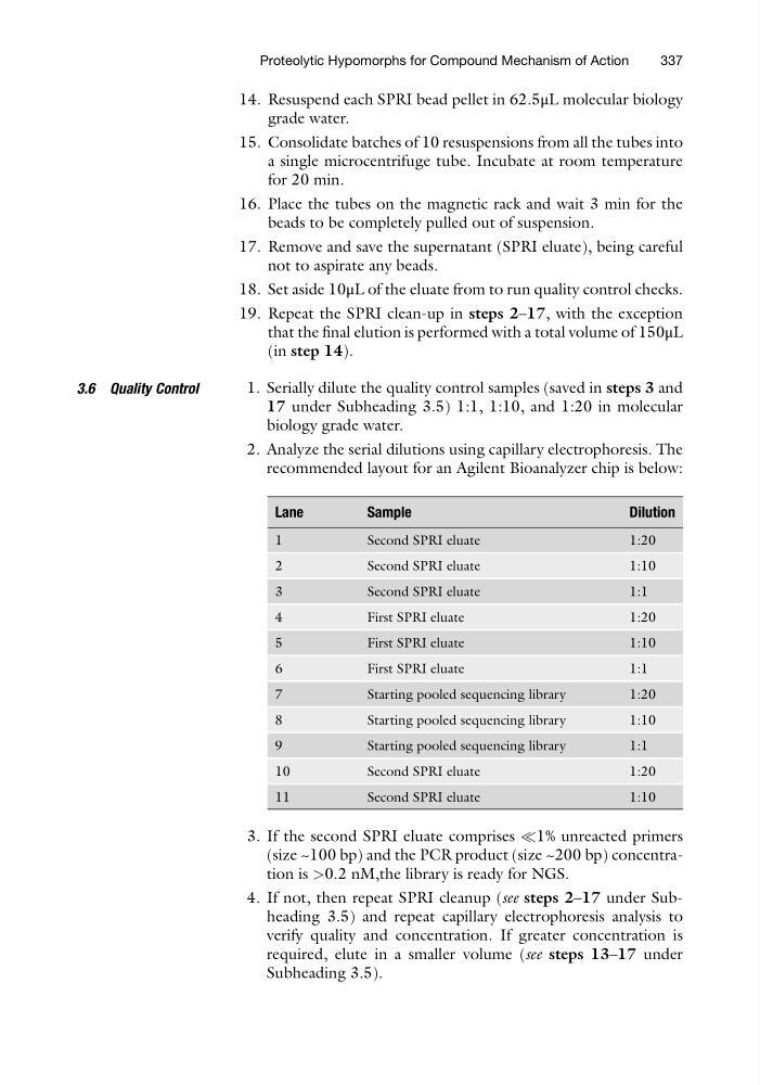
2. Analyze the serial dilutions using capillary electrophoresis. The
recommended layout for an Agilent Bioanalyzer chip is below:
Lane
Sample
Dilution
1
Second SPRI eluate
1:20
2
Second SPRI eluate
1:10
3
Second SPRI eluate
1:1
4
First SPRI eluate
1:20
5
First SPRI eluate
1:10
6
First SPRI eluate
1:1
7
Starting pooled sequencing library
1:20
8
Starting pooled sequencing library
1:10
9
Starting pooled sequencing library
1:1
10
Second SPRI eluate
1:20
11
Second SPRI eluate
1:10
3. If the second SPRI eluate comprises (1% unreacted primers
(size ~100 bp) and the PCR product (size ~200 bp) concentration is >0.2 nM,the library is ready for NGS.
4. If not, then repeat SPRI cleanup (see steps 2–17 under Subheading 3.5) and repeat capillary electrophoresis analysis to
verify quality and concentration. If greater concentration is
required, elute in a smaller volume (see steps 13–17 under
Subheading 3.5).
Proteolytic Hypomorphs for Compound Mechanism of Action
337
grade water.
15. Consolidate batches of 10 resuspensions from all the tubes into
a single microcentrifuge tube. Incubate at room temperature
for 20 min.
16. Place the tubes on the magnetic rack and wait 3 min for the
beads to be completely pulled out of suspension.
17. Remove and save the supernatant (SPRI eluate), being careful
not to aspirate any beads.
18. Set aside 10μL of the eluate from to run quality control checks.
19. Repeat the SPRI clean-up in steps 2–17, with the exception
that the final elution is performed with a total volume of 150μL
(in step 14).
3.6 Quality Control
1. Serially dilute the quality control samples (saved in steps 3 and
17 under Subheading 3.5) 1:1, 1:10, and 1:20 in molecular
biology grade water.
2. Analyze the serial dilutions using capillary electrophoresis. The
recommended layout for an Agilent Bioanalyzer chip is below:
Lane
Sample
Dilution
1
Second SPRI eluate
1:20
2
Second SPRI eluate
1:10
3
Second SPRI eluate
1:1
4
First SPRI eluate
1:20
5
First SPRI eluate
1:10
6
First SPRI eluate
1:1
7
Starting pooled sequencing library
1:20
8
Starting pooled sequencing library
1:10
9
Starting pooled sequencing library
1:1
10
Second SPRI eluate
1:20
11
Second SPRI eluate
1:10
3. If the second SPRI eluate comprises (1% unreacted primers
(size ~100 bp) and the PCR product (size ~200 bp) concentration is >0.2 nM,the library is ready for NGS.
4. If not, then repeat SPRI cleanup (see steps 2–17 under Subheading 3.5) and repeat capillary electrophoresis analysis to
verify quality and concentration. If greater concentration is
required, elute in a smaller volume (see steps 13–17 under
Subheading 3.5).
Proteolytic Hypomorphs for Compound Mechanism of Action
337