

4. Centrifuge fixed mycobacteria at 3000 Â g for at least 5 min
and resuspend in 1–5 mL of Flow Cytometry Buffer (see Note
15).
5. Transfer samples to round-bottom tubes and run flow cytometer according to manufacturer’s instructions and experimental design. Acquire FSC and SSC of mycobacteria using a
logarithmic scale.
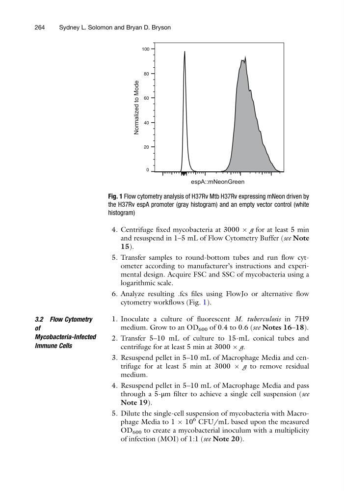
6. Analyze resulting .fcs files using FlowJo or alternative flow
cytometry workflows (Fig. 1).
3.2 Flow Cytometry
of
Mycobacteria-Infected
Immune Cells
1. Inoculate a culture of fluorescent M. tuberculosis in 7H9
medium. Grow to an OD 600 of 0.4 to 0.6 (see Notes 16–18).
2. Transfer 5–10 mL of culture to 15-mL conical tubes and
centrifuge for at least 5 min at 3000 Â g.
3. Resuspend pellet in 5–10 mL of Macrophage Media and centrifuge for at least 5 min at 3000 Â g to remove residual
medium.
4. Resuspend pellet in 5–10 mL of Macrophage Media and pass
through a 5-μm filter to achieve a single cell suspension (see
Note 19).
5. Dilute the single-cell suspension of mycobacteria with Macrophage Media to 1 Â 10
6 CFU/mL based upon the measured
OD 600 to create a mycobacterial inoculum with a multiplicity
of infection (MOI) of 1:1 (see Note 20).
0
20
40
60
80
100
Normalized to Mode
espA::mNeonGreen
Fig. 1 Flow cytometry analysis of H37Rv Mtb H37Rv expressing mNeon driven by
the H37Rv espA promoter (gray histogram) and an empty vector control (white
histogram)
264
Sydney L. Solomon and Bryan D. Bryson
and resuspend in 1–5 mL of Flow Cytometry Buffer (see Note
15).
5. Transfer samples to round-bottom tubes and run flow cytometer according to manufacturer’s instructions and experimental design. Acquire FSC and SSC of mycobacteria using a
logarithmic scale.
6. Analyze resulting .fcs files using FlowJo or alternative flow
cytometry workflows (Fig. 1).
3.2 Flow Cytometry
of
Mycobacteria-Infected
Immune Cells
1. Inoculate a culture of fluorescent M. tuberculosis in 7H9
medium. Grow to an OD 600 of 0.4 to 0.6 (see Notes 16–18).
2. Transfer 5–10 mL of culture to 15-mL conical tubes and
centrifuge for at least 5 min at 3000 Â g.
3. Resuspend pellet in 5–10 mL of Macrophage Media and centrifuge for at least 5 min at 3000 Â g to remove residual
medium.
4. Resuspend pellet in 5–10 mL of Macrophage Media and pass
through a 5-μm filter to achieve a single cell suspension (see
Note 19).
5. Dilute the single-cell suspension of mycobacteria with Macrophage Media to 1 Â 10
6 CFU/mL based upon the measured
OD 600 to create a mycobacterial inoculum with a multiplicity
of infection (MOI) of 1:1 (see Note 20).
0
20
40
60
80
100
Normalized to Mode
espA::mNeonGreen
Fig. 1 Flow cytometry analysis of H37Rv Mtb H37Rv expressing mNeon driven by
the H37Rv espA promoter (gray histogram) and an empty vector control (white
histogram)
264
Sydney L. Solomon and Bryan D. Bryson