

(a) Single-cell segmentation: This procedure is useful to
extract several different parameters from individual cells
over time, such as cell size “Area”; absolute cell fluorescence “Integrated density”; fluorescence intensity normalized to the cell size “Mean gray value”; variation among
pixels “Standard deviation,” etc., which can be selected in
“Set Measurements” of ImageJ. To carry out assisted manual segmentation, open the “ROI Manager” macros and
enable the “Selection Brush Tool” to draw polygons on
individual cells. The width of the brush can be adjusted
according to the cell width, e.g., 7 to 9 pixels for
M. smegmatis and 3 to 5 pixels for M. tuberculosis. To
analyze time-lapse image sequences, the polygon has to
be modified frame by frame as the cell grows over time,
and each new polygon drawn on the profile of a cell at a
given time point is added to the “ROI Manager” list.
Cells can be renamed or numbered to keep track of different cells in the same time-lapse movie. Lineage information can also be documented within the “ROI Manager”
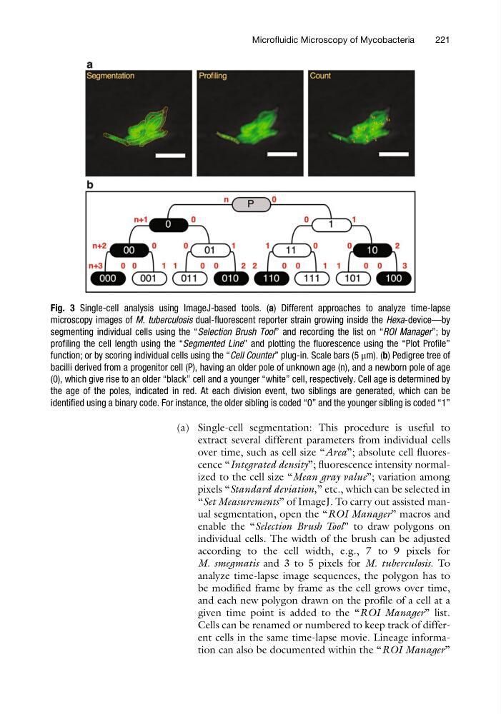
Fig. 3 Single-cell analysis using ImageJ-based tools. (a) Different approaches to analyze time-lapse
microscopy images of M. tuberculosis dual-fluorescent reporter strain growing inside the Hexa-device—by
segmenting individual cells using the “Selection Brush Tool” and recording the list on “ROI Manager”; by
profiling the cell length using the “Segmented Line” and plotting the fluorescence using the “Plot Profile”
function; or by scoring individual cells using the “Cell Counter” plug-in. Scale bars (5 μm). (b) Pedigree tree of
bacilli derived from a progenitor cell (P), having an older pole of unknown age (n), and a newborn pole of age
(0), which give rise to an older “black” cell and a younger “white” cell, respectively. Cell age is determined by
the age of the poles, indicated in red. At each division event, two siblings are generated, which can be
identified using a binary code. For instance, the older sibling is coded “0” and the younger sibling is coded “1”
Microfluidic Microscopy of Mycobacteria
221
extract several different parameters from individual cells
over time, such as cell size “Area”; absolute cell fluorescence “Integrated density”; fluorescence intensity normalized to the cell size “Mean gray value”; variation among
pixels “Standard deviation,” etc., which can be selected in
“Set Measurements” of ImageJ. To carry out assisted manual segmentation, open the “ROI Manager” macros and
enable the “Selection Brush Tool” to draw polygons on
individual cells. The width of the brush can be adjusted
according to the cell width, e.g., 7 to 9 pixels for
M. smegmatis and 3 to 5 pixels for M. tuberculosis. To
analyze time-lapse image sequences, the polygon has to
be modified frame by frame as the cell grows over time,
and each new polygon drawn on the profile of a cell at a
given time point is added to the “ROI Manager” list.
Cells can be renamed or numbered to keep track of different cells in the same time-lapse movie. Lineage information can also be documented within the “ROI Manager”
Fig. 3 Single-cell analysis using ImageJ-based tools. (a) Different approaches to analyze time-lapse
microscopy images of M. tuberculosis dual-fluorescent reporter strain growing inside the Hexa-device—by
segmenting individual cells using the “Selection Brush Tool” and recording the list on “ROI Manager”; by
profiling the cell length using the “Segmented Line” and plotting the fluorescence using the “Plot Profile”
function; or by scoring individual cells using the “Cell Counter” plug-in. Scale bars (5 μm). (b) Pedigree tree of
bacilli derived from a progenitor cell (P), having an older pole of unknown age (n), and a newborn pole of age
(0), which give rise to an older “black” cell and a younger “white” cell, respectively. Cell age is determined by
the age of the poles, indicated in red. At each division event, two siblings are generated, which can be
identified using a binary code. For instance, the older sibling is coded “0” and the younger sibling is coded “1”
Microfluidic Microscopy of Mycobacteria
221