

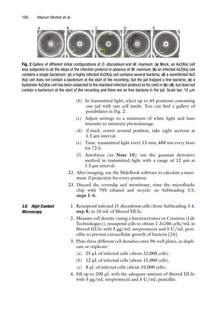
(b) In transmitted light, select up to 45 positions containing
one jail with one cell inside. You can find a gallery of
possibilities in Fig. 2.
(c) Adjust settings to a minimum of white light and laser
intensity to minimize photodamage.
(d) Z-stack: center around position, take eight sections at
1.5 μm interval.
(e) Time: transmitted light every 15 min; 488 nm every hour
for 72 h.
(f) Autofocus (see Note 10): use the gaussian derivative
method in transmitted light with a range of 12 μm at
1.5 μm interval.
22. After imaging, use the SlideBook software to calculate a maximum Z projection for every position.
23. Discard the coverslip and membrane, rinse the microfluidic
chip with 70% ethanol and recycle: see Subheading 3.5,
steps 1–6.
3.6 High-Content
Microscopy
1. Resuspend infected D. discoideum cells (from Subheading 3.4,
step 8) in 10 mL of filtered HL5c.
2. Measure cell density (using a haemocytomer or Countess (Life
Technologies)), resuspend cells to obtain 1.3x10⁶ cells/mL in
filtered HL5c with 5 μg/mL streptomycin and 5 U/mL penicillin to prevent extracellular growth of bacteria [24].
3. Plate three different cell densities onto 96-well plates, in duplicate or triplicate:
(a) 25 μL of infected cells (about 32,000 cells).
(b) 12 μL of infected cells (about 15,000 cells).
(c) 8 μL of infected cells (about 10,000 cells).
4. Fill up to 200 μL with the adequate amount of filtered HL5c
with 5 μg/mL streptomycin and 5 U/mL penicillin.
Fig. 2 Gallery of different initial configurations of D. discoideum and M. marinum. (a) Mock, an Ax2(Ka) cell
was subjected to all the steps of the infection protocol in absence of M. marinum; (b) an infected Ax2(Ka) cell
contains a single bacterium; (c) a highly infected Ax2(Ka) cell contains several bacteria; (d) a noninfected Ax2
(Ka) cell does not contain a bacterium at the start of the recording, but the jail trapped a few bacteria; (e) a
bystander Ax2(Ka) cell has been subjected to the standard infection protocol as for cells in (b)–(d), but does not
contain a bacterium at the start of the recording and there are no free bacteria in the jail. Scale bar: 10 μm
192
Manon Mottet et al.
one jail with one cell inside. You can find a gallery of
possibilities in Fig. 2.
(c) Adjust settings to a minimum of white light and laser
intensity to minimize photodamage.
(d) Z-stack: center around position, take eight sections at
1.5 μm interval.
(e) Time: transmitted light every 15 min; 488 nm every hour
for 72 h.
(f) Autofocus (see Note 10): use the gaussian derivative
method in transmitted light with a range of 12 μm at
1.5 μm interval.
22. After imaging, use the SlideBook software to calculate a maximum Z projection for every position.
23. Discard the coverslip and membrane, rinse the microfluidic
chip with 70% ethanol and recycle: see Subheading 3.5,
steps 1–6.
3.6 High-Content
Microscopy
1. Resuspend infected D. discoideum cells (from Subheading 3.4,
step 8) in 10 mL of filtered HL5c.
2. Measure cell density (using a haemocytomer or Countess (Life
Technologies)), resuspend cells to obtain 1.3x10⁶ cells/mL in
filtered HL5c with 5 μg/mL streptomycin and 5 U/mL penicillin to prevent extracellular growth of bacteria [24].
3. Plate three different cell densities onto 96-well plates, in duplicate or triplicate:
(a) 25 μL of infected cells (about 32,000 cells).
(b) 12 μL of infected cells (about 15,000 cells).
(c) 8 μL of infected cells (about 10,000 cells).
4. Fill up to 200 μL with the adequate amount of filtered HL5c
with 5 μg/mL streptomycin and 5 U/mL penicillin.
Fig. 2 Gallery of different initial configurations of D. discoideum and M. marinum. (a) Mock, an Ax2(Ka) cell
was subjected to all the steps of the infection protocol in absence of M. marinum; (b) an infected Ax2(Ka) cell
contains a single bacterium; (c) a highly infected Ax2(Ka) cell contains several bacteria; (d) a noninfected Ax2
(Ka) cell does not contain a bacterium at the start of the recording, but the jail trapped a few bacteria; (e) a
bystander Ax2(Ka) cell has been subjected to the standard infection protocol as for cells in (b)–(d), but does not
contain a bacterium at the start of the recording and there are no free bacteria in the jail. Scale bar: 10 μm
192
Manon Mottet et al.