

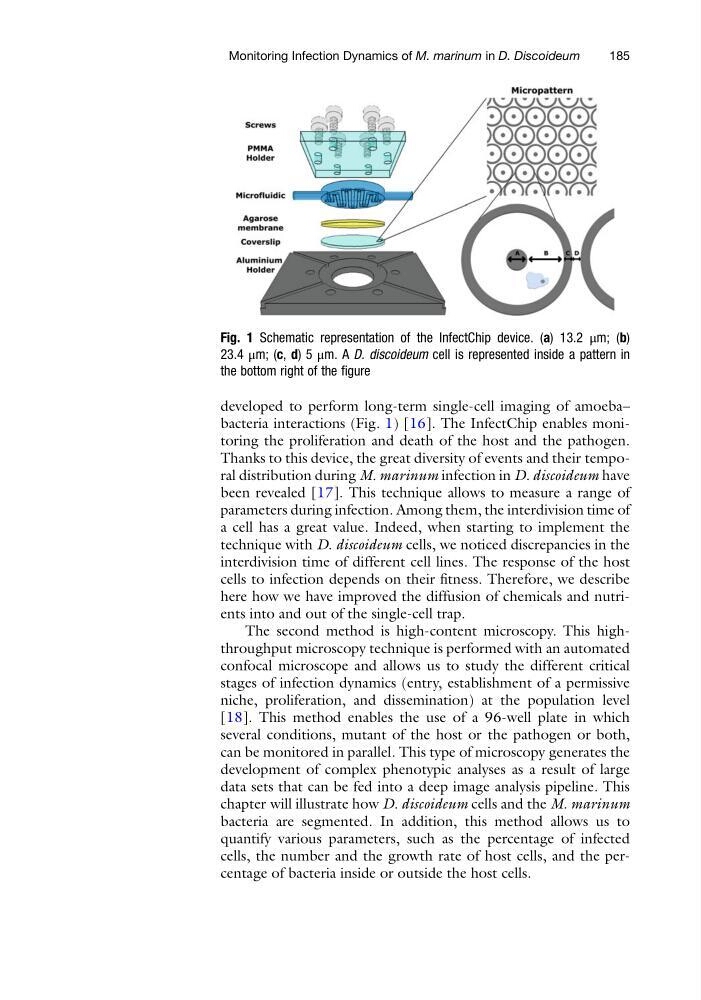
developed to perform long-term single-cell imaging of amoeba–
bacteria interactions (Fig. 1) [16]. The InfectChip enables monitoring the proliferation and death of the host and the pathogen.
Thanks to this device, the great diversity of events and their temporal distribution during M. marinum infection in D. discoideum have
been revealed [17]. This technique allows to measure a range of
parameters during infection. Among them, the interdivision time of
a cell has a great value. Indeed, when starting to implement the
technique with D. discoideum cells, we noticed discrepancies in the
interdivision time of different cell lines. The response of the host
cells to infection depends on their fitness. Therefore, we describe
here how we have improved the diffusion of chemicals and nutrients into and out of the single-cell trap.
The second method is high-content microscopy. This highthroughput microscopy technique is performed with an automated
confocal microscope and allows us to study the different critical
stages of infection dynamics (entry, establishment of a permissive
niche, proliferation, and dissemination) at the population level
[18]. This method enables the use of a 96-well plate in which
several conditions, mutant of the host or the pathogen or both,
can be monitored in parallel. This type of microscopy generates the
development of complex phenotypic analyses as a result of large
data sets that can be fed into a deep image analysis pipeline. This
chapter will illustrate how D. discoideum cells and the M. marinum
bacteria are segmented. In addition, this method allows us to
quantify various parameters, such as the percentage of infected
cells, the number and the growth rate of host cells, and the percentage of bacteria inside or outside the host cells.
Fig. 1 Schematic representation of the InfectChip device. (a) 13.2 μm; (b)
23.4 μm; (c, d) 5 μm. A D. discoideum cell is represented inside a pattern in
the bottom right of the figure
Monitoring Infection Dynamics of M. marinum in D. Discoideum
185
bacteria interactions (Fig. 1) [16]. The InfectChip enables monitoring the proliferation and death of the host and the pathogen.
Thanks to this device, the great diversity of events and their temporal distribution during M. marinum infection in D. discoideum have
been revealed [17]. This technique allows to measure a range of
parameters during infection. Among them, the interdivision time of
a cell has a great value. Indeed, when starting to implement the
technique with D. discoideum cells, we noticed discrepancies in the
interdivision time of different cell lines. The response of the host
cells to infection depends on their fitness. Therefore, we describe
here how we have improved the diffusion of chemicals and nutrients into and out of the single-cell trap.
The second method is high-content microscopy. This highthroughput microscopy technique is performed with an automated
confocal microscope and allows us to study the different critical
stages of infection dynamics (entry, establishment of a permissive
niche, proliferation, and dissemination) at the population level
[18]. This method enables the use of a 96-well plate in which
several conditions, mutant of the host or the pathogen or both,
can be monitored in parallel. This type of microscopy generates the
development of complex phenotypic analyses as a result of large
data sets that can be fed into a deep image analysis pipeline. This
chapter will illustrate how D. discoideum cells and the M. marinum
bacteria are segmented. In addition, this method allows us to
quantify various parameters, such as the percentage of infected
cells, the number and the growth rate of host cells, and the percentage of bacteria inside or outside the host cells.
Fig. 1 Schematic representation of the InfectChip device. (a) 13.2 μm; (b)
23.4 μm; (c, d) 5 μm. A D. discoideum cell is represented inside a pattern in
the bottom right of the figure
Monitoring Infection Dynamics of M. marinum in D. Discoideum
185