

9. Using a hooked syringe needle, draw the coverslip up off the
bottom of the well, and remove with from the well with forceps
(see Note 6). Take care to note which side of the coverslip is
covered with the monolayer. Dip the coverslip into tissue
culture-grade PBS and remove the excess PBS by touching
the side of the coverslip onto a sheet of paper towel.
10. Place the coverslip onto the microscope slide containing the
ProLong
® Gold antifade reagent, monolayer side down. Avoid
capturing air bubbles under the glass. If bubbles are observed,
very gently press on the coverslip to push air bubbles to the
edge of the coverslip.
11. Allow the ProLong
® Gold antifade reagent to cure for 48 h in a
biological safety cabinet.
12. Seal the coverslip to the microscope slide by painting the edges
of the coverslip and immediately surrounding microscope slide
with a clear, fast drying nail polish.
13. Once dry, decontaminate the slides by submerging them in a
10% bleach solution for 10 min or other approved decontamination procedure and remove materials from the BSL-3
laboratory.
4 Notes
1. When preparing tissue culture reagents be sure to use
endotoxin-free plasticware to avoid activation of the macrophages. Measure reagents using disposable plastic pipettes or,
for larger volumes sterile, 50 mL conical tubes or the volume
gradations on the top portion of the filtration unit.
2. To prepare L-cell conditioned medium, quickly thaw a tube of
L929 fibroblasts (ATCC) at 37
C. Gently pipette thawed cells
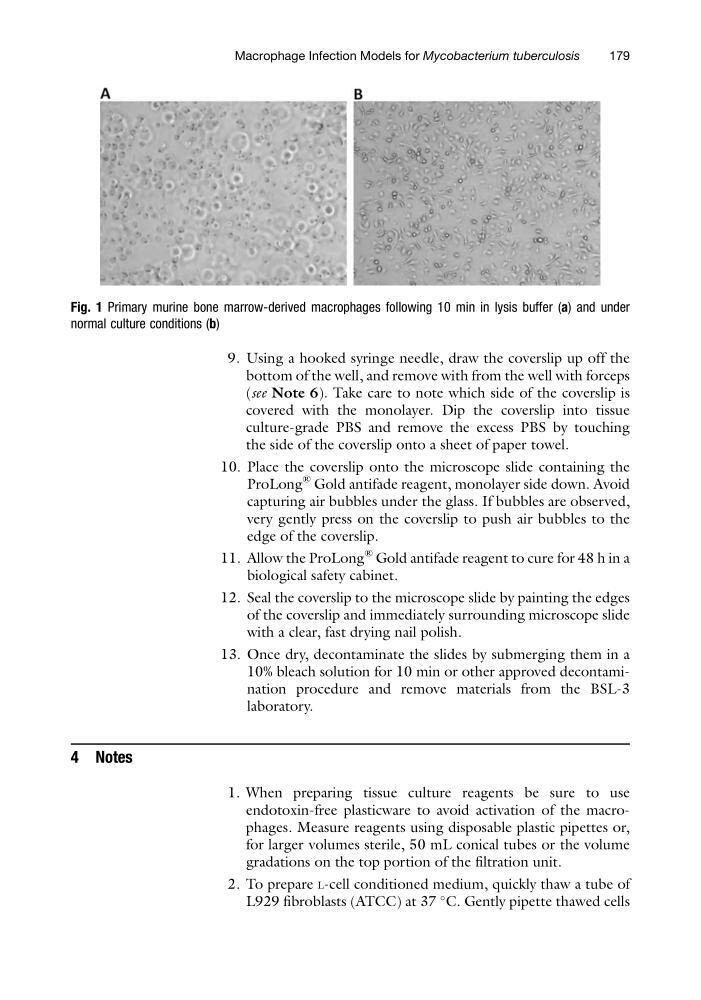
Fig. 1 Primary murine bone marrow-derived macrophages following 10 min in lysis buffer (a) and under
normal culture conditions (b)
Macrophage Infection Models for Mycobacterium tuberculosis
179
bottom of the well, and remove with from the well with forceps
(see Note 6). Take care to note which side of the coverslip is
covered with the monolayer. Dip the coverslip into tissue
culture-grade PBS and remove the excess PBS by touching
the side of the coverslip onto a sheet of paper towel.
10. Place the coverslip onto the microscope slide containing the
ProLong
® Gold antifade reagent, monolayer side down. Avoid
capturing air bubbles under the glass. If bubbles are observed,
very gently press on the coverslip to push air bubbles to the
edge of the coverslip.
11. Allow the ProLong
® Gold antifade reagent to cure for 48 h in a
biological safety cabinet.
12. Seal the coverslip to the microscope slide by painting the edges
of the coverslip and immediately surrounding microscope slide
with a clear, fast drying nail polish.
13. Once dry, decontaminate the slides by submerging them in a
10% bleach solution for 10 min or other approved decontamination procedure and remove materials from the BSL-3
laboratory.
4 Notes
1. When preparing tissue culture reagents be sure to use
endotoxin-free plasticware to avoid activation of the macrophages. Measure reagents using disposable plastic pipettes or,
for larger volumes sterile, 50 mL conical tubes or the volume
gradations on the top portion of the filtration unit.
2. To prepare L-cell conditioned medium, quickly thaw a tube of
L929 fibroblasts (ATCC) at 37
C. Gently pipette thawed cells
Fig. 1 Primary murine bone marrow-derived macrophages following 10 min in lysis buffer (a) and under
normal culture conditions (b)
Macrophage Infection Models for Mycobacterium tuberculosis
179