

3. Vortex the mixture and sonicate for 5 min.
4. Add 1 μL of the internal standard (from step 1) and 8 μL of the
mycolic acid mixture (from step 2) in a 1.5 mL vial equipped
with a glass insert.
5. Complete to 100 μL by adding the base mix (90% isopropanol,
10% acetonitrile, 0.1% formic acid (v/v/v) (see item 8 in Subheading 2.7).
6. Vortex.
7. Sonicate for 5 min before the injection.
8. Equilibrate the system with 80% of the solvent A and 20% of the
solvent B at a rate of 100 μL/min.
9. Inject 5 μL of sample in the column.
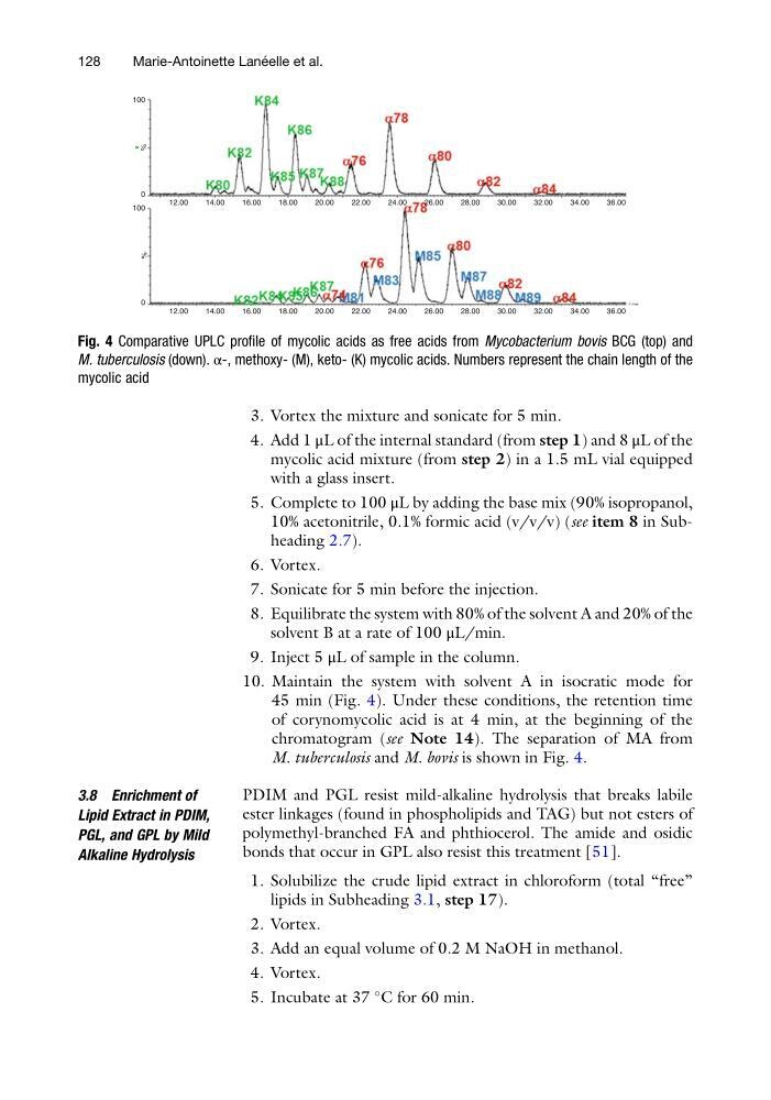
10. Maintain the system with solvent A in isocratic mode for
45 min (Fig. 4). Under these conditions, the retention time
of corynomycolic acid is at 4 min, at the beginning of the
chromatogram (see Note 14). The separation of MA from
M. tuberculosis and M. bovis is shown in Fig. 4.
3.8 Enrichment of
Lipid Extract in PDIM,
PGL, and GPL by Mild
Alkaline Hydrolysis
PDIM and PGL resist mild-alkaline hydrolysis that breaks labile
ester linkages (found in phospholipids and TAG) but not esters of
polymethyl-branched FA and phthiocerol. The amide and osidic
bonds that occur in GPL also resist this treatment [51].
1. Solubilize the crude lipid extract in chloroform (total “free”
lipids in Subheading 3.1, step 17).
2. Vortex.
3. Add an equal volume of 0.2 M NaOH in methanol.
4. Vortex.
5. Incubate at 37
C for 60 min.
100
0
%
100
0
12.00
14.00
16.00
18.00
20.00
22.00
24.00
26.00
28.00
30.00
32.00
34.00
36.00
12.00
14.00
16.00
18.00
20.00
22.00
24.00
26.00
28.00
30.00
32.00
34.00
36.00
%
Fig. 4 Comparative UPLC profile of mycolic acids as free acids from Mycobacterium bovis BCG (top) and
M. tuberculosis (down). α-, methoxy- (M), keto- (K) mycolic acids. Numbers represent the chain length of the
mycolic acid
128
Marie-Antoinette Lane ´ elle et al.
4. Add 1 μL of the internal standard (from step 1) and 8 μL of the
mycolic acid mixture (from step 2) in a 1.5 mL vial equipped
with a glass insert.
5. Complete to 100 μL by adding the base mix (90% isopropanol,
10% acetonitrile, 0.1% formic acid (v/v/v) (see item 8 in Subheading 2.7).
6. Vortex.
7. Sonicate for 5 min before the injection.
8. Equilibrate the system with 80% of the solvent A and 20% of the
solvent B at a rate of 100 μL/min.
9. Inject 5 μL of sample in the column.
10. Maintain the system with solvent A in isocratic mode for
45 min (Fig. 4). Under these conditions, the retention time
of corynomycolic acid is at 4 min, at the beginning of the
chromatogram (see Note 14). The separation of MA from
M. tuberculosis and M. bovis is shown in Fig. 4.
3.8 Enrichment of
Lipid Extract in PDIM,
PGL, and GPL by Mild
Alkaline Hydrolysis
PDIM and PGL resist mild-alkaline hydrolysis that breaks labile
ester linkages (found in phospholipids and TAG) but not esters of
polymethyl-branched FA and phthiocerol. The amide and osidic
bonds that occur in GPL also resist this treatment [51].
1. Solubilize the crude lipid extract in chloroform (total “free”
lipids in Subheading 3.1, step 17).
2. Vortex.
3. Add an equal volume of 0.2 M NaOH in methanol.
4. Vortex.
5. Incubate at 37
C for 60 min.
100
0
%
100
0
12.00
14.00
16.00
18.00
20.00
22.00
24.00
26.00
28.00
30.00
32.00
34.00
36.00
12.00
14.00
16.00
18.00
20.00
22.00
24.00
26.00
28.00
30.00
32.00
34.00
36.00
%
Fig. 4 Comparative UPLC profile of mycolic acids as free acids from Mycobacterium bovis BCG (top) and
M. tuberculosis (down). α-, methoxy- (M), keto- (K) mycolic acids. Numbers represent the chain length of the
mycolic acid
128
Marie-Antoinette Lane ´ elle et al.