

3.5 Fracturing
of Whole
Mycobacterium spp.
Cells by Bead Beating
(See Note 56)
1. Dilute live or inactivated Mycobacterium spp. cells (see Subheading 3.1) in breaking buffer to a concentration of 0.5 g/
mL based on wet weight of cells (see Note 57).
2. Aliquot 0.5–0.75 mL of the cell suspension into Pre-Filled
Lysing Matrix B tubes and tighten screw cap (see Note 58).
3. Turn on Mini-Bead Beater and set speed to 48 (see Note 59).
4. Place tubes into Mini-Bead Beater, close lid and press START.
Beat for 30–45 s.
5. Remove lysing tube from Mini-Bead Beater and immediately
place on ice for 90 s (see Note 60).
6. Repeat steps 4 and 5 for a total of 10–12 cycles (see Note 61).
7. Place lysis tubes in centrifuge and spin at 1500 Â g for 5 min to
spin down silica beads and generate a semi-cleared lysate (see
Note 62).
8. Remove semi-cleared lysate into sterile 1.5 mL microcentrifuge
tube and centrifuge at 8000 Â g to further remove cellular
debris (see Note 63, for additional steps if breaking live cells).
9. Store cell lysate samples at À80
C.
3.6 Subcellular
Fractionation
of Mycobacterium spp.
Whole-Cell Lysate
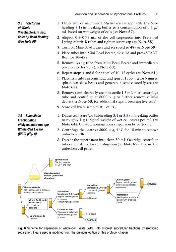
(WCL) (Fig. 6)
1. Dilute cell lysate (see Subheading 3.4 or 3.5) in breaking buffer
to roughly 1 g (original weight of wet cell paste) per mL (see
Note 64). Create a homogenous suspension by vortexing.
2. Centrifuge the lysate at 3000 Â g, 4
C for 15 min to remove
unbroken cells.
3. Decant the supernatant into clean 50 mL Oakridge centrifuge
tubes and balance for centrifugation (see Note 65). Discard the
unbroken cell pellet.
Spent Filtrate
Starting material
for CFP and MEV
Harvested cells
Cell paste used to produce
subcellular fractions
Unclarified
Membrane & Cytosol
Repeat centrifugation
to remove
contaminating cell wall
Unclarified
Membrane & Cytosol
Ultracentrifugation to
separate
Crude Cytosol
Repeat centrifugation to
remove contaminating
membrane
Membrane
Wash pellet surface
gently with breaking
buffer
Cytosol
Whole Cell Lysate
Keep as final
product, or
separate
Unbroken cells
Discard
Crude Cell Wall
Resuspend pellet and
repeat centrifugation
to wash
Cell Wall
Discard
Discard
Mycobacterium
culture (described
elsewhere)
Fig. 6 Scheme for separation of whole-cell lysate (WCL) into discreet subcellular fractions by isopycnic
separation. Figure used is modified from the previous edition of this protocol chapter
Extraction and Separation of Mycobacterial Proteins
93
of Whole
Mycobacterium spp.
Cells by Bead Beating
(See Note 56)
1. Dilute live or inactivated Mycobacterium spp. cells (see Subheading 3.1) in breaking buffer to a concentration of 0.5 g/
mL based on wet weight of cells (see Note 57).
2. Aliquot 0.5–0.75 mL of the cell suspension into Pre-Filled
Lysing Matrix B tubes and tighten screw cap (see Note 58).
3. Turn on Mini-Bead Beater and set speed to 48 (see Note 59).
4. Place tubes into Mini-Bead Beater, close lid and press START.
Beat for 30–45 s.
5. Remove lysing tube from Mini-Bead Beater and immediately
place on ice for 90 s (see Note 60).
6. Repeat steps 4 and 5 for a total of 10–12 cycles (see Note 61).
7. Place lysis tubes in centrifuge and spin at 1500 Â g for 5 min to
spin down silica beads and generate a semi-cleared lysate (see
Note 62).
8. Remove semi-cleared lysate into sterile 1.5 mL microcentrifuge
tube and centrifuge at 8000 Â g to further remove cellular
debris (see Note 63, for additional steps if breaking live cells).
9. Store cell lysate samples at À80
C.
3.6 Subcellular
Fractionation
of Mycobacterium spp.
Whole-Cell Lysate
(WCL) (Fig. 6)
1. Dilute cell lysate (see Subheading 3.4 or 3.5) in breaking buffer
to roughly 1 g (original weight of wet cell paste) per mL (see
Note 64). Create a homogenous suspension by vortexing.
2. Centrifuge the lysate at 3000 Â g, 4
C for 15 min to remove
unbroken cells.
3. Decant the supernatant into clean 50 mL Oakridge centrifuge
tubes and balance for centrifugation (see Note 65). Discard the
unbroken cell pellet.
Spent Filtrate
Starting material
for CFP and MEV
Harvested cells
Cell paste used to produce
subcellular fractions
Unclarified
Membrane & Cytosol
Repeat centrifugation
to remove
contaminating cell wall
Unclarified
Membrane & Cytosol
Ultracentrifugation to
separate
Crude Cytosol
Repeat centrifugation to
remove contaminating
membrane
Membrane
Wash pellet surface
gently with breaking
buffer
Cytosol
Whole Cell Lysate
Keep as final
product, or
separate
Unbroken cells
Discard
Crude Cell Wall
Resuspend pellet and
repeat centrifugation
to wash
Cell Wall
Discard
Discard
Mycobacterium
culture (described
elsewhere)
Fig. 6 Scheme for separation of whole-cell lysate (WCL) into discreet subcellular fractions by isopycnic
separation. Figure used is modified from the previous edition of this protocol chapter
Extraction and Separation of Mycobacterial Proteins
93