

Figure 11.3
226
Bacterial Cellulose
(a)
(b)
(c)
(d)
(e)
(f )
(g)
(h)
(i)
(j)
(k)
(l)
(m)
(n)
(o)
(p)
Figure 11.3
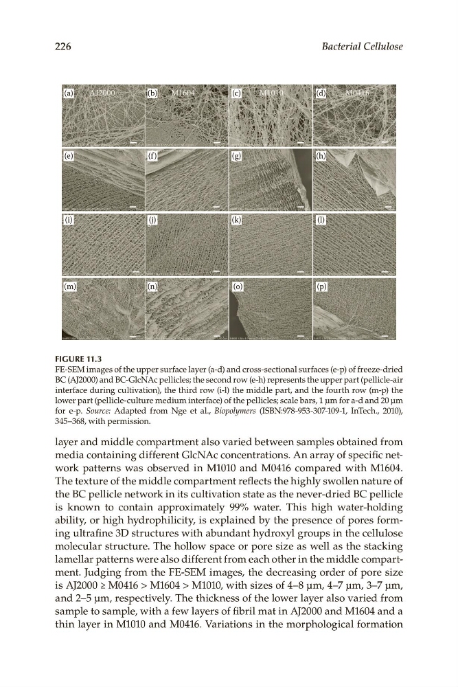
FE-SEM.images.of.the.upper.surface.layer.(a-d).and.cross-sectional.surfaces.(e-p).of.freeze-dried.
BC.(AJ2000).and.BC-GlcNAc.pellicles;.the.second.row.(e-h).represents.the.upper.part.(pellicle-air.
interface.during.cultivation),.the.third.row.(i-l).the.middle.part,.and.the.fourth.row.(m-p).the.
lower.part.(pellicle-culture.medium.interface).of.the.pellicles;.scale.bars,.1.μm.for.a-d.and.20 μm.
for. e-p.. Source:. Adapted. from. Nge. et. al.,. Biopolymers. (ISBN:978-953-307-109-1,. InTech.,. 2010),.
345–368,.with.permission.
layer.and.middle.compartment.also.varied.between.samples.obtained.from.
media.containing.different.GlcNAc.concentrations..An.array.of.specific.network. patterns. was. observed. in. M1010. and. M0416. compared. with. M1604..
The.texture.of.the.middle.compartment.reflects.the.highly.swollen.nature.of.
the.BC.pellicle.network.in.its.cultivation.state.as.the.never-dried.BC.. pellicle.
is. known. to. contain. approximately. 99%. water.. This. high. water-holding.
ability,.or.high.hydrophilicity,.is.explained.by.the.presence.of.pores.forming.ultrafine.3D.structures.with.abundant.hydroxyl.groups.in.the.cellulose.
molecular.structure..The.hollow.space.or.pore.size.as.well.as.the.stacking.
lamellar.patterns.were.also.different.from.each.other.in.the.middle.compartment..Judging.from.the.FE-SEM.images,.the.decreasing.order.of.pore.size.
is.AJ2000.≥.M0416.>.M1604.>.M1010,.with.sizes.of.4–8.μm,.4–7.μm,.3–7.μm,.
and.2–5 μm,.respectively..The.thickness.of.the.lower.layer.also.varied.from.
sample.to.sample,.with.a.few.layers.of.fibril.mat.in.AJ2000.and.M1604.and.a.
thin.layer.in.M1010.and.M0416..Variations.in.the.morphological.formation.
226
Bacterial Cellulose
(a)
(b)
(c)
(d)
(e)
(f )
(g)
(h)
(i)
(j)
(k)
(l)
(m)
(n)
(o)
(p)
Figure 11.3
FE-SEM.images.of.the.upper.surface.layer.(a-d).and.cross-sectional.surfaces.(e-p).of.freeze-dried.
BC.(AJ2000).and.BC-GlcNAc.pellicles;.the.second.row.(e-h).represents.the.upper.part.(pellicle-air.
interface.during.cultivation),.the.third.row.(i-l).the.middle.part,.and.the.fourth.row.(m-p).the.
lower.part.(pellicle-culture.medium.interface).of.the.pellicles;.scale.bars,.1.μm.for.a-d.and.20 μm.
for. e-p.. Source:. Adapted. from. Nge. et. al.,. Biopolymers. (ISBN:978-953-307-109-1,. InTech.,. 2010),.
345–368,.with.permission.
layer.and.middle.compartment.also.varied.between.samples.obtained.from.
media.containing.different.GlcNAc.concentrations..An.array.of.specific.network. patterns. was. observed. in. M1010. and. M0416. compared. with. M1604..
The.texture.of.the.middle.compartment.reflects.the.highly.swollen.nature.of.
the.BC.pellicle.network.in.its.cultivation.state.as.the.never-dried.BC.. pellicle.
is. known. to. contain. approximately. 99%. water.. This. high. water-holding.
ability,.or.high.hydrophilicity,.is.explained.by.the.presence.of.pores.forming.ultrafine.3D.structures.with.abundant.hydroxyl.groups.in.the.cellulose.
molecular.structure..The.hollow.space.or.pore.size.as.well.as.the.stacking.
lamellar.patterns.were.also.different.from.each.other.in.the.middle.compartment..Judging.from.the.FE-SEM.images,.the.decreasing.order.of.pore.size.
is.AJ2000.≥.M0416.>.M1604.>.M1010,.with.sizes.of.4–8.μm,.4–7.μm,.3–7.μm,.
and.2–5 μm,.respectively..The.thickness.of.the.lower.layer.also.varied.from.
sample.to.sample,.with.a.few.layers.of.fibril.mat.in.AJ2000.and.M1604.and.a.
thin.layer.in.M1010.and.M0416..Variations.in.the.morphological.formation.