

Figure 6.12
Figure 6.13
cellulose microfibrils
Driving force
M a c r o m o le c u la r t r a c k s o n N O C
M o v in g d ir e c ti o n
~1 µm
~100 nm
Normal
On the Tracks of NOC
Secreted 3.5 nm
Random
movements
Bacterium
A ribbon
composed of a few hundreds microfibrils
132
Bacterial Cellulose
(a)
(b)
0.00
0.00
500 µm
500 µm
348.7 (nm)
34.1 (nm)
0.0 (nm)
0.0 (nm)
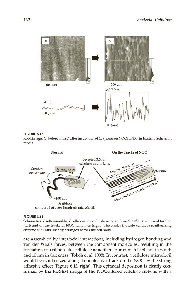
Figure 6.12
AFM.images.(a).before.and.(b).after.incubation.of.G. xylinus.on.NOC.for.10.h.in.Hestrin–Schramm.
media.
Figure 6.13
Schematics.of.self-assembly.of.cellulose.microfibrils.secreted.from.G. xylinus.in.normal.fashion.
(left).and.on.the.tracks.of.NOC.templates.(right)..The.circles.indicate.cellulose-synthesizing.
enzyme.subunits.linearly.arranged.across.the.cell.body.
are.assembled.by.interfacial.interactions,.including.hydrogen.bonding.and.
van. der. Waals. forces,. between. the. component. molecules,. resulting. in. the.
formation.of.a.ribbon-like.cellulose.nanofiber.approximately.50.nm.in.width.
and.10.nm.in.thickness.(Tokoh.et.al..1998)..In.contrast,.a.cellulose.microfibril.
would.be.synthesized.along.the.molecular.track.on.the.NOC.by.the.strong.
adhesive. effect. (Figure 6.13,. right).. This. epitaxial. deposition. is. clearly. confirmed. by. the. FE-SEM. image. of. the. NOC-altered. cellulose. ribbons. with. a.
Figure 6.13
cellulose microfibrils
Driving force
M a c r o m o le c u la r t r a c k s o n N O C
M o v in g d ir e c ti o n
~1 µm
~100 nm
Normal
On the Tracks of NOC
Secreted 3.5 nm
Random
movements
Bacterium
A ribbon
composed of a few hundreds microfibrils
132
Bacterial Cellulose
(a)
(b)
0.00
0.00
500 µm
500 µm
348.7 (nm)
34.1 (nm)
0.0 (nm)
0.0 (nm)
Figure 6.12
AFM.images.(a).before.and.(b).after.incubation.of.G. xylinus.on.NOC.for.10.h.in.Hestrin–Schramm.
media.
Figure 6.13
Schematics.of.self-assembly.of.cellulose.microfibrils.secreted.from.G. xylinus.in.normal.fashion.
(left).and.on.the.tracks.of.NOC.templates.(right)..The.circles.indicate.cellulose-synthesizing.
enzyme.subunits.linearly.arranged.across.the.cell.body.
are.assembled.by.interfacial.interactions,.including.hydrogen.bonding.and.
van. der. Waals. forces,. between. the. component. molecules,. resulting. in. the.
formation.of.a.ribbon-like.cellulose.nanofiber.approximately.50.nm.in.width.
and.10.nm.in.thickness.(Tokoh.et.al..1998)..In.contrast,.a.cellulose.microfibril.
would.be.synthesized.along.the.molecular.track.on.the.NOC.by.the.strong.
adhesive. effect. (Figure 6.13,. right).. This. epitaxial. deposition. is. clearly. confirmed. by. the. FE-SEM. image. of. the. NOC-altered. cellulose. ribbons. with. a.