

53
Signaling Pathways in AP Patterning
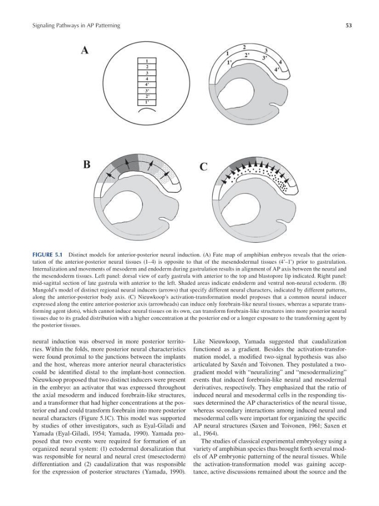
FIGURE 5.1 Distinct models for anterior-posterior neural induction. (A) Fate map of amphibian embryos reveals that the orientation of the anterior-posterior neural tissues (1–4) is opposite to that of the mesendodermal tissues (4’–1’) prior to gastrulation.
Internalization and movements of mesoderm and endoderm during gastrulation results in alignment of AP axis between the neural and
the mesendoderm tissues. Left panel: dorsal view of early gastrula with anterior to the top and blastopore lip indicated. Right panel:
mid-sagittal section of late gastrula with anterior to the left. Shaded areas indicate endoderm and ventral non-neural ectoderm. (B)
Mangold’s model of distinct regional neural inducers (arrows) that specify different neural characters, indicated by different patterns,
along the anterior-posterior body axis. (C) Nieuwkoop’s activation-transformation model proposes that a common neural inducer
expressed along the entire anterior-posterior axis (arrowheads) can induce only forebrain-like neural tissues, whereas a separate transforming agent (dots), which cannot induce neural tissues on its own, can transform forebrain-like structures into more posterior neural
tissues due to its graded distribution with a higher concentration at the posterior end or a longer exposure to the transforming agent by
the posterior tissues.
neural induction was observed in more posterior territories. Within the folds, more posterior neural characteristics
were found proximal to the junctions between the implants
and the host, whereas more anterior neural characteristics
could be identifed distal to the implant-host connection.
Nieuwkoop proposed that two distinct inducers were present
in the embryo: an activator that was expressed throughout
the axial mesoderm and induced forebrain-like structures,
and a transformer that had higher concentrations at the posterior end and could transform forebrain into more posterior
neural characters (Figure 5.1C). This model was supported
by studies of other investigators, such as Eyal-Giladi and
Yamada (Eyal-Giladi, 1954; Yamada, 1990). Yamada proposed that two events were required for formation of an
organized neural system: (1) ectodermal dorsalization that
was responsible for neural and neural crest (mesectoderm)
differentiation and (2) caudalization that was responsible
for the expression of posterior structures (Yamada, 1990).
Like Nieuwkoop, Yamada suggested that caudalization
functioned as a gradient. Besides the activation-transformation model, a modifed two-signal hypothesis was also
articulated by Saxén and Toivonen. They postulated a twogradient model with “neuralizing” and “mesodermalizing”
events that induced forebrain-like neural and mesodermal
derivatives, respectively. They emphasized that the ratio of
induced neural and mesodermal cells in the responding tissues determined the AP characteristics of the neural tissue,
whereas secondary interactions among induced neural and
mesodermal cells were important for organizing the specif c
AP neural structures (Saxen and Toivonen, 1961; Saxen et
al., 1964).
The studies of classical experimental embryology using a
variety of amphibian species thus brought forth several models of AP embryonic patterning of the neural tissues. While
the activation-transformation model was gaining acceptance, active discussions remained about the source and the
Signaling Pathways in AP Patterning
FIGURE 5.1 Distinct models for anterior-posterior neural induction. (A) Fate map of amphibian embryos reveals that the orientation of the anterior-posterior neural tissues (1–4) is opposite to that of the mesendodermal tissues (4’–1’) prior to gastrulation.
Internalization and movements of mesoderm and endoderm during gastrulation results in alignment of AP axis between the neural and
the mesendoderm tissues. Left panel: dorsal view of early gastrula with anterior to the top and blastopore lip indicated. Right panel:
mid-sagittal section of late gastrula with anterior to the left. Shaded areas indicate endoderm and ventral non-neural ectoderm. (B)
Mangold’s model of distinct regional neural inducers (arrows) that specify different neural characters, indicated by different patterns,
along the anterior-posterior body axis. (C) Nieuwkoop’s activation-transformation model proposes that a common neural inducer
expressed along the entire anterior-posterior axis (arrowheads) can induce only forebrain-like neural tissues, whereas a separate transforming agent (dots), which cannot induce neural tissues on its own, can transform forebrain-like structures into more posterior neural
tissues due to its graded distribution with a higher concentration at the posterior end or a longer exposure to the transforming agent by
the posterior tissues.
neural induction was observed in more posterior territories. Within the folds, more posterior neural characteristics
were found proximal to the junctions between the implants
and the host, whereas more anterior neural characteristics
could be identifed distal to the implant-host connection.
Nieuwkoop proposed that two distinct inducers were present
in the embryo: an activator that was expressed throughout
the axial mesoderm and induced forebrain-like structures,
and a transformer that had higher concentrations at the posterior end and could transform forebrain into more posterior
neural characters (Figure 5.1C). This model was supported
by studies of other investigators, such as Eyal-Giladi and
Yamada (Eyal-Giladi, 1954; Yamada, 1990). Yamada proposed that two events were required for formation of an
organized neural system: (1) ectodermal dorsalization that
was responsible for neural and neural crest (mesectoderm)
differentiation and (2) caudalization that was responsible
for the expression of posterior structures (Yamada, 1990).
Like Nieuwkoop, Yamada suggested that caudalization
functioned as a gradient. Besides the activation-transformation model, a modifed two-signal hypothesis was also
articulated by Saxén and Toivonen. They postulated a twogradient model with “neuralizing” and “mesodermalizing”
events that induced forebrain-like neural and mesodermal
derivatives, respectively. They emphasized that the ratio of
induced neural and mesodermal cells in the responding tissues determined the AP characteristics of the neural tissue,
whereas secondary interactions among induced neural and
mesodermal cells were important for organizing the specif c
AP neural structures (Saxen and Toivonen, 1961; Saxen et
al., 1964).
The studies of classical experimental embryology using a
variety of amphibian species thus brought forth several models of AP embryonic patterning of the neural tissues. While
the activation-transformation model was gaining acceptance, active discussions remained about the source and the