

46
Xenopus
the dorsal organizer gives rise not only to notochord and
foor plate but also to the medial somite that lies next to the
notochord. Lineage tracing at the 32-cell stage has delineated in detail the origin of the dorsal lip of the circular
blastopore at early neurula (Bauer et al., 1994; Vodicka and
Gerhart, 1995). When lineage traced at late gastrula/early
neurula, the dorsal lip organizer continues its gastrulation
movements inside the tailbud embryo all the way to the tip
of the tail, where it gives rise to the stem cells of the chordoneural hinge (Gont et al., 1993).
The main fnding of the Spemann organizer molecular
studies was that this tissue is a source of secreted growth
factor antagonists, many of which were novel proteins at the
time ( Figure 4.1A ). Thus, Chordin, Noggin, and Follistatin
were BMP antagonists ( Piccolo et al., 1996 ; Zimmerman et
al., 1996 ; Fainsod et al., 1997 ). The head inducer Cerberus
is a secreted inhibitor of Nodal, BMP, and Wnt ( Piccolo et al.,
1999 ). Dickkopf-1 (Dkk1) ( Glinka et al., 1998 ), Frzb-1,
Crescent, Angptl4, Pkdcc, and Bighead are all secreted Wnt
antagonists ( Figure 4.1A ).
In the case of BMP antagonists, the simultaneous depletion
of all three is required for the loss of all dorsal structures
(Khokha et al., 2005). The depletion of Chordin leads to
a partial loss of dorsal structures and the complete loss of
inductive activity of transplanted organizers (Oelgeschläger
et al., 2003). Activin, a TGF-β superfamily growth factor that
induces dorsal mesoderm in animal cap ectodermal explants,
is only able to induce ventral mesoderm when Chordin is
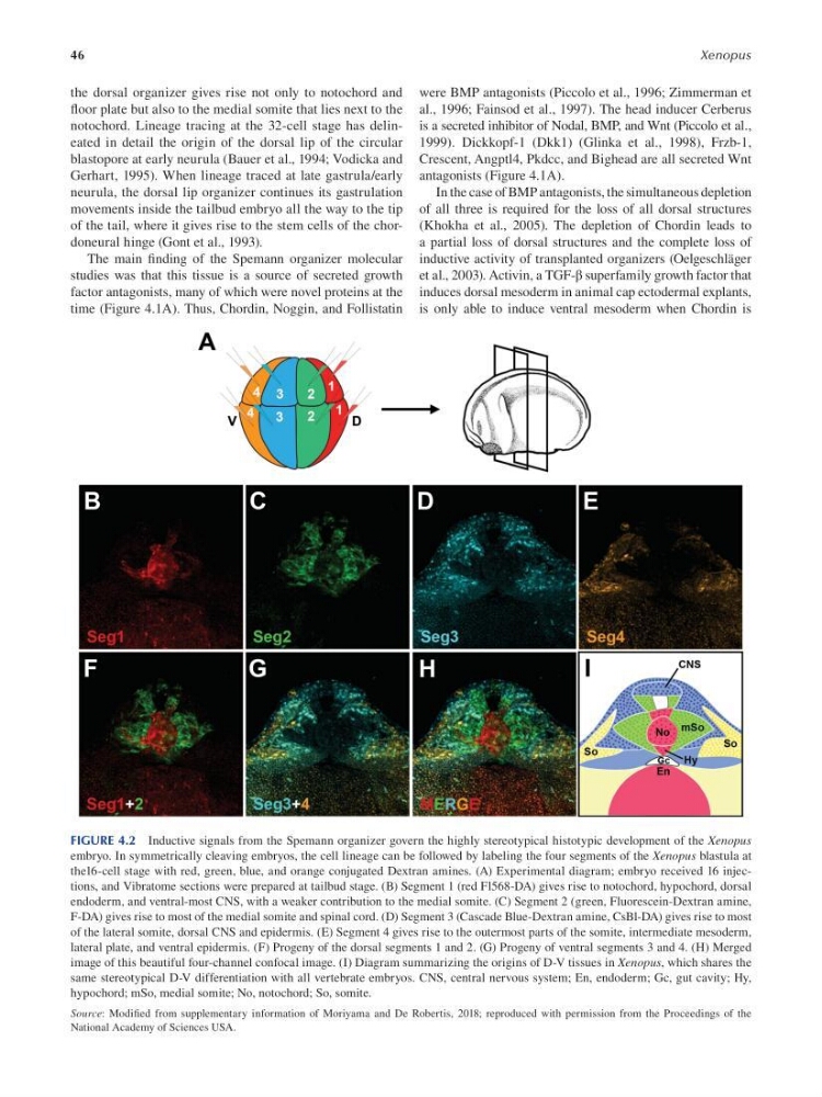
FIGURE 4.2 Inductive signals from the Spemann organizer govern the highly stereotypical histotypic development of the Xenopus
embryo. In symmetrically cleaving embryos, the cell lineage can be followed by labeling the four segments of the Xenopus blastula at
the16-cell stage with red, green, blue, and orange conjugated Dextran amines. (A) Experimental diagram; embryo received 16 injections, and Vibratome sections were prepared at tailbud stage. (B) Segment 1 (red Fl568-DA) gives rise to notochord, hypochord, dorsal
endoderm, and ventral-most CNS, with a weaker contribution to the medial somite. (C) Segment 2 (green, Fluorescein-Dextran amine,
F-DA) gives rise to most of the medial somite and spinal cord. (D) Segment 3 (Cascade Blue-Dextran amine, CsBl-DA) gives rise to most
of the lateral somite, dorsal CNS and epidermis. (E) Segment 4 gives rise to the outermost parts of the somite, intermediate mesoderm,
lateral plate, and ventral epidermis. (F) Progeny of the dorsal segments 1 and 2. (G) Progeny of ventral segments 3 and 4. (H) Merged
image of this beautiful four-channel confocal image. (I) Diagram summarizing the origins of D-V tissues in Xenopus, which shares the
same stereotypical D-V differentiation with all vertebrate embryos. CNS, central nervous system; En, endoderm; Gc, gut cavity; Hy,
hypochord; mSo, medial somite; No, notochord; So, somite.
Source: Modifed from supplementary information of Moriyama and De Robertis, 2018; reproduced with permission from the Proceedings of the
National Academy of Sciences USA.
Xenopus
the dorsal organizer gives rise not only to notochord and
foor plate but also to the medial somite that lies next to the
notochord. Lineage tracing at the 32-cell stage has delineated in detail the origin of the dorsal lip of the circular
blastopore at early neurula (Bauer et al., 1994; Vodicka and
Gerhart, 1995). When lineage traced at late gastrula/early
neurula, the dorsal lip organizer continues its gastrulation
movements inside the tailbud embryo all the way to the tip
of the tail, where it gives rise to the stem cells of the chordoneural hinge (Gont et al., 1993).
The main fnding of the Spemann organizer molecular
studies was that this tissue is a source of secreted growth
factor antagonists, many of which were novel proteins at the
time ( Figure 4.1A ). Thus, Chordin, Noggin, and Follistatin
were BMP antagonists ( Piccolo et al., 1996 ; Zimmerman et
al., 1996 ; Fainsod et al., 1997 ). The head inducer Cerberus
is a secreted inhibitor of Nodal, BMP, and Wnt ( Piccolo et al.,
1999 ). Dickkopf-1 (Dkk1) ( Glinka et al., 1998 ), Frzb-1,
Crescent, Angptl4, Pkdcc, and Bighead are all secreted Wnt
antagonists ( Figure 4.1A ).
In the case of BMP antagonists, the simultaneous depletion
of all three is required for the loss of all dorsal structures
(Khokha et al., 2005). The depletion of Chordin leads to
a partial loss of dorsal structures and the complete loss of
inductive activity of transplanted organizers (Oelgeschläger
et al., 2003). Activin, a TGF-β superfamily growth factor that
induces dorsal mesoderm in animal cap ectodermal explants,
is only able to induce ventral mesoderm when Chordin is
FIGURE 4.2 Inductive signals from the Spemann organizer govern the highly stereotypical histotypic development of the Xenopus
embryo. In symmetrically cleaving embryos, the cell lineage can be followed by labeling the four segments of the Xenopus blastula at
the16-cell stage with red, green, blue, and orange conjugated Dextran amines. (A) Experimental diagram; embryo received 16 injections, and Vibratome sections were prepared at tailbud stage. (B) Segment 1 (red Fl568-DA) gives rise to notochord, hypochord, dorsal
endoderm, and ventral-most CNS, with a weaker contribution to the medial somite. (C) Segment 2 (green, Fluorescein-Dextran amine,
F-DA) gives rise to most of the medial somite and spinal cord. (D) Segment 3 (Cascade Blue-Dextran amine, CsBl-DA) gives rise to most
of the lateral somite, dorsal CNS and epidermis. (E) Segment 4 gives rise to the outermost parts of the somite, intermediate mesoderm,
lateral plate, and ventral epidermis. (F) Progeny of the dorsal segments 1 and 2. (G) Progeny of ventral segments 3 and 4. (H) Merged
image of this beautiful four-channel confocal image. (I) Diagram summarizing the origins of D-V tissues in Xenopus, which shares the
same stereotypical D-V differentiation with all vertebrate embryos. CNS, central nervous system; En, endoderm; Gc, gut cavity; Hy,
hypochord; mSo, medial somite; No, notochord; So, somite.
Source: Modifed from supplementary information of Moriyama and De Robertis, 2018; reproduced with permission from the Proceedings of the
National Academy of Sciences USA.