

27
Maternal mRNAs and Cell Lineages
and undergoes specifc localization to the vegetal cortex of
full-grown oocytes, becoming inherited by vegetal blastomeres ( Weeks and Melton, 1987 ). Tangential efforts to
clone maternally expressed Wingless-type MMTV integration site (Wnt) genes fortuitously identifed an additional
vegetally localized mRNA, Xwnt-11 (now wnt11b; Ku and
Melton, 1993 ). Both the Tgf β and Wnt proteins had been
concomitantly implicated in mesoderm induction and in axis
formation (see Section 4 ), making the presence of these proteins in the set of vegetally localized transcripts especially
intriguing.
Further fractionation of the oocyte using biochemical or
physical methods facilitated the isolation of additional localized mRNAs (Elinson et al., 1993; Pondel and King, 1988)
and led to the demonstration of two mechanisms of mRNA
localization in the oocyte: (1) a late pathway initiated after
the elaboration of animal-vegetal polarity in mid-oogenesis and (2) an early mechanism in pre-vitellogenic oocytes
involving the mitochondrial cloud (Forristall et al., 1995;
Kloc and Etkin, 1995). The early pattern exactly matched
the known distribution of the germ plasm in Xenopus
(Czołowska, 1972, 1969; Heasman et al., 1984; Savage and
Danilchik, 1993). An intermediate pattern also became
evident following the characterization of plin2 (née fatvg)
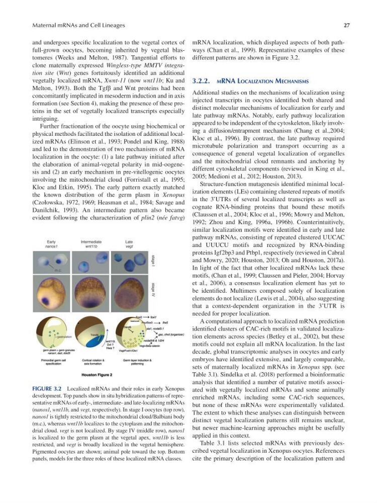
FIGURE 3.2 Localized mRNAs and their roles in early Xenopus
development. Top panels show in situ hybridization patterns of representative mRNAs of early-, intermediate- and late-localizing mRNAs
(nanos1, wnt11b, and vegt, respectively). In stage I oocytes (top row),
nanos1 is tightly restricted to the mitochondrial cloud/Balbiani body
(m.c.), whereas wnt11b localizes to the cytoplasm and the mitochondrial cloud. vegt is not localized. By stage IV (middle row), nanos1
is localized to the germ plasm at the vegetal apex, wnt11b is less
restricted, and vegt is broadly localized in the vegetal hemisphere.
Pigmented oocytes are shown; animal pole toward the top. Bottom
panels, models for the three roles of these localized mRNA classes.
mRNA localization, which displayed aspects of both pathways (Chan et al., 1999). Representative examples of these
different patterns are shown in Figure 3.2.
3.2.2. MRNA LOCALIZATION MECHANISMS
Additional studies on the mechanisms of localization using
injected transcripts in oocytes identifed both shared and
distinct molecular mechanisms of localization for early and
late pathway mRNAs. Notably, early pathway localization
appeared to be independent of the cytoskeleton, likely involving a diffusion/entrapment mechanism (Chang et al.,2004;
Kloc et al., 1996 ). By contrast, the late pathway required
microtubule polarization and transport occurring as a
consequence of general vegetal localization of organelles
and the mitochondrial cloud remnants and anchoring by
different cytoskeletal components (reviewed in King et al.,
2005; Medioni et al., 2012; Houston, 2013).
Structure-function mutagenesis identifed minimal localization elements (LEs) containing clustered repeats of motifs
in the 3’UTRs of several localized transcripts as well as
cognate RNA-binding proteins that bound these motifs
(Claussen et al., 2004; Kloc et al., 1996; Mowry and Melton,
1992; Zhou and King, 1996a, 1996b). Counterintuitively,
similar localization motifs were identifed in early and late
pathway mRNAs, consisting of repeated clustered UUCAC
and UUUCU motifs and recognized by RNA-binding
proteins Igf2bp3 and Ptbp1, respectively (reviewed in Cabral
and Mowry, 2020; Houston, 2013; Oh and Houston, 2017a).
In light of the fact that other localized mRNAs lack these
motifs, (Chan et al., 1999; Claussen and Pieler, 2004; Horvay
et al., 2006), a consensus localization element has yet to
be identifed. Multimers composed solely of localization
elements do not localize (Lewis et al., 2004), also suggesting
that a context-dependent organization in the 3’UTR is
needed for proper localization.
A computational approach to localized mRNA prediction
identifed clusters of CAC-rich motifs in validated localization elements across species (Betley et al., 2002), but these
motifs could not explain all mRNA localization. In the last
decade, global transcriptomic analyses in oocytes and early
embryos have identifed extensive, and largely comparable,
sets of maternally localized mRNAs in Xenopus spp. (see
Table 3.1). Sindelka et al. (2018) performed a bioinformatic
analysis that identifed a number of putative motifs associated with vegetally localized mRNAs and some animally
enriched mRNAs, including some CAC-rich sequences,
but none of these mRNAs were experimentally validated.
The extent to which these analyses can distinguish between
distinct vegetal localization patterns still remains unclear,
but newer machine-learning approaches might be usefully
applied in this context.
Table 3.1 lists selected mRNAs with previously described vegetal localization in Xenopus oocytes. References
cite the primary description of the localization pattern and
Maternal mRNAs and Cell Lineages
and undergoes specifc localization to the vegetal cortex of
full-grown oocytes, becoming inherited by vegetal blastomeres ( Weeks and Melton, 1987 ). Tangential efforts to
clone maternally expressed Wingless-type MMTV integration site (Wnt) genes fortuitously identifed an additional
vegetally localized mRNA, Xwnt-11 (now wnt11b; Ku and
Melton, 1993 ). Both the Tgf β and Wnt proteins had been
concomitantly implicated in mesoderm induction and in axis
formation (see Section 4 ), making the presence of these proteins in the set of vegetally localized transcripts especially
intriguing.
Further fractionation of the oocyte using biochemical or
physical methods facilitated the isolation of additional localized mRNAs (Elinson et al., 1993; Pondel and King, 1988)
and led to the demonstration of two mechanisms of mRNA
localization in the oocyte: (1) a late pathway initiated after
the elaboration of animal-vegetal polarity in mid-oogenesis and (2) an early mechanism in pre-vitellogenic oocytes
involving the mitochondrial cloud (Forristall et al., 1995;
Kloc and Etkin, 1995). The early pattern exactly matched
the known distribution of the germ plasm in Xenopus
(Czołowska, 1972, 1969; Heasman et al., 1984; Savage and
Danilchik, 1993). An intermediate pattern also became
evident following the characterization of plin2 (née fatvg)
FIGURE 3.2 Localized mRNAs and their roles in early Xenopus
development. Top panels show in situ hybridization patterns of representative mRNAs of early-, intermediate- and late-localizing mRNAs
(nanos1, wnt11b, and vegt, respectively). In stage I oocytes (top row),
nanos1 is tightly restricted to the mitochondrial cloud/Balbiani body
(m.c.), whereas wnt11b localizes to the cytoplasm and the mitochondrial cloud. vegt is not localized. By stage IV (middle row), nanos1
is localized to the germ plasm at the vegetal apex, wnt11b is less
restricted, and vegt is broadly localized in the vegetal hemisphere.
Pigmented oocytes are shown; animal pole toward the top. Bottom
panels, models for the three roles of these localized mRNA classes.
mRNA localization, which displayed aspects of both pathways (Chan et al., 1999). Representative examples of these
different patterns are shown in Figure 3.2.
3.2.2. MRNA LOCALIZATION MECHANISMS
Additional studies on the mechanisms of localization using
injected transcripts in oocytes identifed both shared and
distinct molecular mechanisms of localization for early and
late pathway mRNAs. Notably, early pathway localization
appeared to be independent of the cytoskeleton, likely involving a diffusion/entrapment mechanism (Chang et al.,2004;
Kloc et al., 1996 ). By contrast, the late pathway required
microtubule polarization and transport occurring as a
consequence of general vegetal localization of organelles
and the mitochondrial cloud remnants and anchoring by
different cytoskeletal components (reviewed in King et al.,
2005; Medioni et al., 2012; Houston, 2013).
Structure-function mutagenesis identifed minimal localization elements (LEs) containing clustered repeats of motifs
in the 3’UTRs of several localized transcripts as well as
cognate RNA-binding proteins that bound these motifs
(Claussen et al., 2004; Kloc et al., 1996; Mowry and Melton,
1992; Zhou and King, 1996a, 1996b). Counterintuitively,
similar localization motifs were identifed in early and late
pathway mRNAs, consisting of repeated clustered UUCAC
and UUUCU motifs and recognized by RNA-binding
proteins Igf2bp3 and Ptbp1, respectively (reviewed in Cabral
and Mowry, 2020; Houston, 2013; Oh and Houston, 2017a).
In light of the fact that other localized mRNAs lack these
motifs, (Chan et al., 1999; Claussen and Pieler, 2004; Horvay
et al., 2006), a consensus localization element has yet to
be identifed. Multimers composed solely of localization
elements do not localize (Lewis et al., 2004), also suggesting
that a context-dependent organization in the 3’UTR is
needed for proper localization.
A computational approach to localized mRNA prediction
identifed clusters of CAC-rich motifs in validated localization elements across species (Betley et al., 2002), but these
motifs could not explain all mRNA localization. In the last
decade, global transcriptomic analyses in oocytes and early
embryos have identifed extensive, and largely comparable,
sets of maternally localized mRNAs in Xenopus spp. (see
Table 3.1). Sindelka et al. (2018) performed a bioinformatic
analysis that identifed a number of putative motifs associated with vegetally localized mRNAs and some animally
enriched mRNAs, including some CAC-rich sequences,
but none of these mRNAs were experimentally validated.
The extent to which these analyses can distinguish between
distinct vegetal localization patterns still remains unclear,
but newer machine-learning approaches might be usefully
applied in this context.
Table 3.1 lists selected mRNAs with previously described vegetal localization in Xenopus oocytes. References
cite the primary description of the localization pattern and