

266
Xenopus
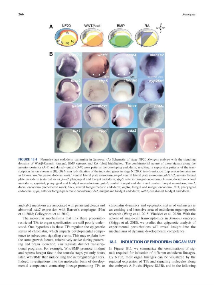
FIGURE 18.4 Neurula-stage endoderm patterning in Xenopus. (A) Schematic of stage NF20 Xenopus embryo with the signaling
domains of Wnt/β-Catenin (orange), BMP (green), and RA (blue) highlighted. The combinatorial nature of these signals along the
anterior-posterior (A-P) and dorsal-ventral (D-V) axes patterns the developing endoderm, resulting in expression patterns of the transcription factors shown in (B). (B) In situ hybridization of the indicated genes in stage NF20 X. laevis embryos. Expression domains are
as follows: sox17a, pan-endoderm; wnt11, ventral lateral plate mesoderm; bmp4, ventral lateral plate mesoderm; aldh1a2, anterior lateral
plate mesoderm (external view); foxa2, pharyngeal and foregut endoderm; sfrp5, anterior foregut endoderm; chordin, dorsal notochord
mesoderm; cyp26a1, pharyngeal and hindgut mesendoderm; gata6, ventral foregut endoderm and ventral foregut mesoderm; mnx1,
dorsal endoderm (archenteron roof); hhex, ventral foregut/hepatic endoderm; hnf4α, foregut and midgut endoderm; tbx1, pharyngeal
endoderm; vpp1, anterior foregut/pancreatic endoderm; cdx2, midgut and hindgut endoderm; satb2, distal-most hindgut endoderm.
and cdx2 mutations are associated with persistent cloaca and
abnormal cdx2 expression with Barrett’s esophagus (Hsu
et al. 2018; Colleypriest et al. 2010).
The molecular mechanisms that link these progenitorrestricted TFs to organ specifcation are still poorly understood. One hypothesis is these TFs regulate the epigenetic
status of chromatin, which imparts developmental competence to subsequent signaling events. This may explain how
the same growth factors, reiteratively active during patterning and organ induction, can regulate distinct transcriptional programs. For example, Wnt/BMP promote hindgut
and repress foregut fate in the neurula stage, yet only hours
later, Wnt/BMP then induce lung fate in foregut progenitors.
Indeed, investigations into the molecular basis of developmental competence connecting lineage-promoting TFs to
chromatin dynamics and epigenetic status of enhancers is
an exciting and intensive area of endoderm organogenesis
research (Wang et al. 2015; Vinckier et al. 2020). With the
advent of single-cell transcriptomics in Xenopus embryos
(Briggs et al. 2018), we predict that epigenetic analysis of
experimental perturbations will reveal insight into the
mechanisms of dynamic developmental competence.
18.5. INDUCTION OF ENDODERM ORGAN FATE
In Figure 18.5, we summarize the combinations of signals required for induction of different endoderm lineages.
By NF35, most organ lineages can be visualized by the
regional expression of TFs and signaling molecules along
the embryo’s A-P axis (Figure 18.5B), and in the following
Xenopus
FIGURE 18.4 Neurula-stage endoderm patterning in Xenopus. (A) Schematic of stage NF20 Xenopus embryo with the signaling
domains of Wnt/β-Catenin (orange), BMP (green), and RA (blue) highlighted. The combinatorial nature of these signals along the
anterior-posterior (A-P) and dorsal-ventral (D-V) axes patterns the developing endoderm, resulting in expression patterns of the transcription factors shown in (B). (B) In situ hybridization of the indicated genes in stage NF20 X. laevis embryos. Expression domains are
as follows: sox17a, pan-endoderm; wnt11, ventral lateral plate mesoderm; bmp4, ventral lateral plate mesoderm; aldh1a2, anterior lateral
plate mesoderm (external view); foxa2, pharyngeal and foregut endoderm; sfrp5, anterior foregut endoderm; chordin, dorsal notochord
mesoderm; cyp26a1, pharyngeal and hindgut mesendoderm; gata6, ventral foregut endoderm and ventral foregut mesoderm; mnx1,
dorsal endoderm (archenteron roof); hhex, ventral foregut/hepatic endoderm; hnf4α, foregut and midgut endoderm; tbx1, pharyngeal
endoderm; vpp1, anterior foregut/pancreatic endoderm; cdx2, midgut and hindgut endoderm; satb2, distal-most hindgut endoderm.
and cdx2 mutations are associated with persistent cloaca and
abnormal cdx2 expression with Barrett’s esophagus (Hsu
et al. 2018; Colleypriest et al. 2010).
The molecular mechanisms that link these progenitorrestricted TFs to organ specifcation are still poorly understood. One hypothesis is these TFs regulate the epigenetic
status of chromatin, which imparts developmental competence to subsequent signaling events. This may explain how
the same growth factors, reiteratively active during patterning and organ induction, can regulate distinct transcriptional programs. For example, Wnt/BMP promote hindgut
and repress foregut fate in the neurula stage, yet only hours
later, Wnt/BMP then induce lung fate in foregut progenitors.
Indeed, investigations into the molecular basis of developmental competence connecting lineage-promoting TFs to
chromatin dynamics and epigenetic status of enhancers is
an exciting and intensive area of endoderm organogenesis
research (Wang et al. 2015; Vinckier et al. 2020). With the
advent of single-cell transcriptomics in Xenopus embryos
(Briggs et al. 2018), we predict that epigenetic analysis of
experimental perturbations will reveal insight into the
mechanisms of dynamic developmental competence.
18.5. INDUCTION OF ENDODERM ORGAN FATE
In Figure 18.5, we summarize the combinations of signals required for induction of different endoderm lineages.
By NF35, most organ lineages can be visualized by the
regional expression of TFs and signaling molecules along
the embryo’s A-P axis (Figure 18.5B), and in the following