

14
Xenopus
extract from Xenopus eggs and showed that somatic frog
nuclei added to it could be induced to undergo DNA replication. This experiment confrmed that nuclei from cells
in the quiescent state (liver) could be induced to enter S
phase by egg cytosol and that DNA replication would occur
by recruitment of necessary components from the extract.
These experiments supported the notion that the major drivers of cell cycle progression were in fact cytoplasmic factors
and not nuclear ones.
A critical breakthrough in egg extract preparation and
use was made by Masui and Lohka in 1983. Using concentrated cytoplasmic extract prepared from Rana pipiens eggs
and sperm nuclei from Xenopus laevis, they documented
morphological and biochemical changes associated with
progression through the cell cycle. These events included
nuclear assembly, DNA replication, and after some time
had elapsed, nuclear envelope breakdown and chromosome
condensation. Importantly, in this experiment, the nuclei
had been pre-treated with detergent to remove membranes
and many nuclear-associated proteins. The results suggested
that the nuclei had assembled and progressed through the
cell cycle through the recruitment of components from the
extract. Through fractionation of the extract, they were further able to show that both the soluble and particulate components of the extract were required for DNA replication
to occur: the particulate component contained membrane
vesicles that could fuse to form a nuclear envelope, and the
soluble fraction contained proteins and other factors, such as
nucleotides, critical for nuclear assembly and function.
In experiments related to those using egg extracts,
Gerhart, Kirschner, and colleagues characterized an activity
which they named maturation promoting factor (MPF) using
Xenopus eggs and oocytes. MPF was defned as a cytoplasmic activity that could be transferred from a mature egg into
a late-staged oocyte, causing the recipient oocyte to mature
into an egg (Masui and Markert, 1971; Smith and Ecker,
1971). The fact that during maturation, the meiotic cell cycle
is driven from meiosis I to arrest into meiosis II suggested
that MPF, a cytoplasmic factor, might itself be a driver of
the cell cycle. MPF had striking and unusual properties: it
caused oocyte maturation even when signif cantly diluted
(100-fold!) prior to injection into the recipient oocyte, and
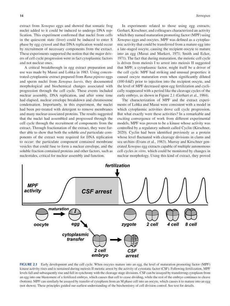
the level of MPF decreased upon egg fertilization and cyclically reappeared with a period like the cleavage cycles of the
early embryo, as shown in Figure 2.1 (Gerhart et al., 1984).
The characterization of MPF and the extract experiments of Lohka and Masui were consistent with a model in
which cytoplasmic activities drove cell cycle progression.
But what exactly were these activities? In a remarkable and
exciting convergence of work from different experimental
models, MPF was proven to be a kinase whose activity was
controlled by a regulatory subunit called Cyclin ( Kirschner,
2020 ). Cyclin had been identifed previously as a protein
whose level f uctuated with cleavage divisions in clams and
sea urchins ( Evans et al., 1983 ). Murray and Kirschner generated Xenopus egg extracts capable of multiple autonomous
cell cycles in vitro, which could be monitored by changes in
nuclear morphology. Using this kind of extract, they proved
FIGURE 2.1 Early development and the cell cycle. When oocytes mature into an egg, the level of maturation promoting factor (MPF)
kinase activity rises and is sustained during meiosis II meiotic arrest by the activity of cytostatic factor (CSF). Following fertilization, MPF
levels fall and subsequently rise and fall in synchrony with the cleavage stage divisions. CSF can be assayed by transferring cytoplasm from
an egg into one blastomere of a fertilized embryo, which causes that cell to cease dividing, while the rest of the embryo continues to cleave
(bottom). MPF can similarly be assayed by transfer of cytoplasm from an M phase cell into an oocyte, which causes it to mature into an egg
(not shown). These principles guided our earliest understanding of the biochemistry of cell division control. See text for details.
Xenopus
extract from Xenopus eggs and showed that somatic frog
nuclei added to it could be induced to undergo DNA replication. This experiment confrmed that nuclei from cells
in the quiescent state (liver) could be induced to enter S
phase by egg cytosol and that DNA replication would occur
by recruitment of necessary components from the extract.
These experiments supported the notion that the major drivers of cell cycle progression were in fact cytoplasmic factors
and not nuclear ones.
A critical breakthrough in egg extract preparation and
use was made by Masui and Lohka in 1983. Using concentrated cytoplasmic extract prepared from Rana pipiens eggs
and sperm nuclei from Xenopus laevis, they documented
morphological and biochemical changes associated with
progression through the cell cycle. These events included
nuclear assembly, DNA replication, and after some time
had elapsed, nuclear envelope breakdown and chromosome
condensation. Importantly, in this experiment, the nuclei
had been pre-treated with detergent to remove membranes
and many nuclear-associated proteins. The results suggested
that the nuclei had assembled and progressed through the
cell cycle through the recruitment of components from the
extract. Through fractionation of the extract, they were further able to show that both the soluble and particulate components of the extract were required for DNA replication
to occur: the particulate component contained membrane
vesicles that could fuse to form a nuclear envelope, and the
soluble fraction contained proteins and other factors, such as
nucleotides, critical for nuclear assembly and function.
In experiments related to those using egg extracts,
Gerhart, Kirschner, and colleagues characterized an activity
which they named maturation promoting factor (MPF) using
Xenopus eggs and oocytes. MPF was defned as a cytoplasmic activity that could be transferred from a mature egg into
a late-staged oocyte, causing the recipient oocyte to mature
into an egg (Masui and Markert, 1971; Smith and Ecker,
1971). The fact that during maturation, the meiotic cell cycle
is driven from meiosis I to arrest into meiosis II suggested
that MPF, a cytoplasmic factor, might itself be a driver of
the cell cycle. MPF had striking and unusual properties: it
caused oocyte maturation even when signif cantly diluted
(100-fold!) prior to injection into the recipient oocyte, and
the level of MPF decreased upon egg fertilization and cyclically reappeared with a period like the cleavage cycles of the
early embryo, as shown in Figure 2.1 (Gerhart et al., 1984).
The characterization of MPF and the extract experiments of Lohka and Masui were consistent with a model in
which cytoplasmic activities drove cell cycle progression.
But what exactly were these activities? In a remarkable and
exciting convergence of work from different experimental
models, MPF was proven to be a kinase whose activity was
controlled by a regulatory subunit called Cyclin ( Kirschner,
2020 ). Cyclin had been identifed previously as a protein
whose level f uctuated with cleavage divisions in clams and
sea urchins ( Evans et al., 1983 ). Murray and Kirschner generated Xenopus egg extracts capable of multiple autonomous
cell cycles in vitro, which could be monitored by changes in
nuclear morphology. Using this kind of extract, they proved
FIGURE 2.1 Early development and the cell cycle. When oocytes mature into an egg, the level of maturation promoting factor (MPF)
kinase activity rises and is sustained during meiosis II meiotic arrest by the activity of cytostatic factor (CSF). Following fertilization, MPF
levels fall and subsequently rise and fall in synchrony with the cleavage stage divisions. CSF can be assayed by transferring cytoplasm from
an egg into one blastomere of a fertilized embryo, which causes that cell to cease dividing, while the rest of the embryo continues to cleave
(bottom). MPF can similarly be assayed by transfer of cytoplasm from an M phase cell into an oocyte, which causes it to mature into an egg
(not shown). These principles guided our earliest understanding of the biochemistry of cell division control. See text for details.