

179
Chromatin Remodeling during Development
As it turns out, the characteristics observed in Xenopus are
conserved among vertebrates. In zebrafsh, the histone modifcation dynamics appear quite similar to those in Xenopus
(Lindeman et al. 2011). Even in mouse and human preimplantation embryos, which are never completely devoid
of histone modifcations, the permissive H3K4me3 mark
at ZGA precedes a wave of H3K27 methylation (Liu et al.
2016; Xia et al. 2019). Bivalency is also much less frequent
in mouse embryos compared to mouse embryonic stem cells
(Liu et al. 2016).
In cleavage-stage Xenopus embryos, H3K4me3 and
H3K27me3 appear to be absent. It should be noted that
any constitutive chromatin-associated epitope will double
with every cleavage division of the embryo. Therefore, it is
important to address by how much H3K4me3 increases on
a per-cell or per-unit-of-chromatin basis. This is not necessarily resolved in ChIP-sequencing experiments. Even if a
modifcation is hardly present, it is still possible that what
little is present is highly localized in particular genomic
regions, producing highly enriched ChIP-sequencing peaks.
ChIP with quantitative PCR allows quantifying the percentage recovery relative to input DNA for a particular sequence,
which then can be compared across stages. The median
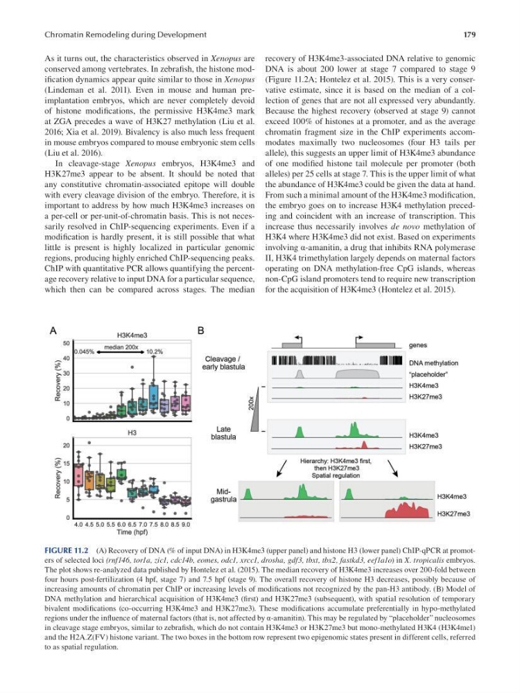
recovery of H3K4me3-associated DNA relative to genomic
DNA is about 200 lower at stage 7 compared to stage 9
(Figure 11.2A; Hontelez et al. 2015). This is a very conservative estimate, since it is based on the median of a collection of genes that are not all expressed very abundantly.
Because the highest recovery (observed at stage 9) cannot
exceed 100% of histones at a promoter, and as the average
chromatin fragment size in the ChIP experiments accommodates maximally two nucleosomes (four H3 tails per
allele), this suggests an upper limit of H3K4me3 abundance
of one modifed histone tail molecule per promoter (both
alleles) per 25 cells at stage 7. This is the upper limit of what
the abundance of H3K4me3 could be given the data at hand.
From such a minimal amount of the H3K4me3 modif cation,
the embryo goes on to increase H3K4 methylation preceding and coincident with an increase of transcription. This
increase thus necessarily involves de novo methylation of
H3K4 where H3K4me3 did not exist. Based on experiments
involving α-amanitin, a drug that inhibits RNA polymerase
II, H3K4 trimethylation largely depends on maternal factors
operating on DNA methylation-free CpG islands, whereas
non-CpG island promoters tend to require new transcription
for the acquisition of H3K4me3 (Hontelez et al. 2015).
FIGURE 11.2 (A) Recovery of DNA (% of input DNA) in H3K4me3 (upper panel) and histone H3 (lower panel) ChIP-qPCR at promoters of selected loci (rnf146, tor1a, zic1, cdc14b, eomes, odc1, xrcc1, drosha, gdf3, tbxt, tbx2, fastkd3, eef1a1o) in X. tropicalis embryos.
The plot shows re-analyzed data published by Hontelez et al. (2015 ). The median recovery of H3K4me3 increases over 200-fold between
four hours post-fertilization (4 hpf, stage 7) and 7.5 hpf (stage 9). The overall recovery of histone H3 decreases, possibly because of
increasing amounts of chromatin per ChIP or increasing levels of modifcations not recognized by the pan-H3 antibody. (B) Model of
DNA methylation and hierarchical acquisition of H3K4me3 (frst) and H3K27me3 (subsequent), with spatial resolution of temporary
bivalent modifcations (co-occurring H3K4me3 and H3K27me3). These modifcations accumulate preferentially in hypo-methylated
regions under the infuence of maternal factors (that is, not affected by α-amanitin). This may be regulated by “placeholder” nucleosomes
in cleavage stage embryos, similar to zebrafsh, which do not contain H3K4me3 or H3K27me3 but mono-methylated H3K4 (H3K4me1)
and the H2A.Z(FV) histone variant. The two boxes in the bottom row represent two epigenomic states present in different cells, referred
to as spatial regulation.
Chromatin Remodeling during Development
As it turns out, the characteristics observed in Xenopus are
conserved among vertebrates. In zebrafsh, the histone modifcation dynamics appear quite similar to those in Xenopus
(Lindeman et al. 2011). Even in mouse and human preimplantation embryos, which are never completely devoid
of histone modifcations, the permissive H3K4me3 mark
at ZGA precedes a wave of H3K27 methylation (Liu et al.
2016; Xia et al. 2019). Bivalency is also much less frequent
in mouse embryos compared to mouse embryonic stem cells
(Liu et al. 2016).
In cleavage-stage Xenopus embryos, H3K4me3 and
H3K27me3 appear to be absent. It should be noted that
any constitutive chromatin-associated epitope will double
with every cleavage division of the embryo. Therefore, it is
important to address by how much H3K4me3 increases on
a per-cell or per-unit-of-chromatin basis. This is not necessarily resolved in ChIP-sequencing experiments. Even if a
modifcation is hardly present, it is still possible that what
little is present is highly localized in particular genomic
regions, producing highly enriched ChIP-sequencing peaks.
ChIP with quantitative PCR allows quantifying the percentage recovery relative to input DNA for a particular sequence,
which then can be compared across stages. The median
recovery of H3K4me3-associated DNA relative to genomic
DNA is about 200 lower at stage 7 compared to stage 9
(Figure 11.2A; Hontelez et al. 2015). This is a very conservative estimate, since it is based on the median of a collection of genes that are not all expressed very abundantly.
Because the highest recovery (observed at stage 9) cannot
exceed 100% of histones at a promoter, and as the average
chromatin fragment size in the ChIP experiments accommodates maximally two nucleosomes (four H3 tails per
allele), this suggests an upper limit of H3K4me3 abundance
of one modifed histone tail molecule per promoter (both
alleles) per 25 cells at stage 7. This is the upper limit of what
the abundance of H3K4me3 could be given the data at hand.
From such a minimal amount of the H3K4me3 modif cation,
the embryo goes on to increase H3K4 methylation preceding and coincident with an increase of transcription. This
increase thus necessarily involves de novo methylation of
H3K4 where H3K4me3 did not exist. Based on experiments
involving α-amanitin, a drug that inhibits RNA polymerase
II, H3K4 trimethylation largely depends on maternal factors
operating on DNA methylation-free CpG islands, whereas
non-CpG island promoters tend to require new transcription
for the acquisition of H3K4me3 (Hontelez et al. 2015).
FIGURE 11.2 (A) Recovery of DNA (% of input DNA) in H3K4me3 (upper panel) and histone H3 (lower panel) ChIP-qPCR at promoters of selected loci (rnf146, tor1a, zic1, cdc14b, eomes, odc1, xrcc1, drosha, gdf3, tbxt, tbx2, fastkd3, eef1a1o) in X. tropicalis embryos.
The plot shows re-analyzed data published by Hontelez et al. (2015 ). The median recovery of H3K4me3 increases over 200-fold between
four hours post-fertilization (4 hpf, stage 7) and 7.5 hpf (stage 9). The overall recovery of histone H3 decreases, possibly because of
increasing amounts of chromatin per ChIP or increasing levels of modifcations not recognized by the pan-H3 antibody. (B) Model of
DNA methylation and hierarchical acquisition of H3K4me3 (frst) and H3K27me3 (subsequent), with spatial resolution of temporary
bivalent modifcations (co-occurring H3K4me3 and H3K27me3). These modifcations accumulate preferentially in hypo-methylated
regions under the infuence of maternal factors (that is, not affected by α-amanitin). This may be regulated by “placeholder” nucleosomes
in cleavage stage embryos, similar to zebrafsh, which do not contain H3K4me3 or H3K27me3 but mono-methylated H3K4 (H3K4me1)
and the H2A.Z(FV) histone variant. The two boxes in the bottom row represent two epigenomic states present in different cells, referred
to as spatial regulation.