

132
Xenopus
contributions to endodermal organs (thymus, thyroid). This
has led to their being considered by some a “fourth germ
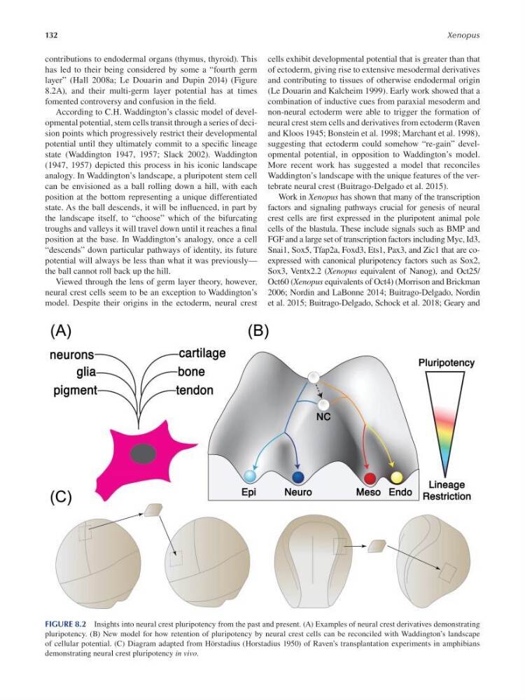
layer” (Hall 2008a; Le Douarin and Dupin 2014) (Figure
8.2A), and their multi-germ layer potential has at times
fomented controversy and confusion in the f eld.
According to C.H. Waddington’s classic model of developmental potential, stem cells transit through a series of decision points which progressively restrict their developmental
potential until they ultimately commit to a specif c lineage
state (Waddington 1947, 1957; Slack 2002 ). Waddington
(1947, 1957 ) depicted this process in his iconic landscape
analogy. In Waddington’s landscape, a pluripotent stem cell
can be envisioned as a ball rolling down a hill, with each
position at the bottom representing a unique differentiated
state. As the ball descends, it will be infuenced, in part by
the landscape itself, to “choose” which of the bifurcating
troughs and valleys it will travel down until it reaches a f nal
position at the base. In Waddington’s analogy, once a cell
“descends” down particular pathways of identity, its future
potential will always be less than what it was previously—
the ball cannot roll back up the hill.
Viewed through the lens of germ layer theory, however,
neural crest cells seem to be an exception to Waddington’s
model. Despite their origins in the ectoderm, neural crest
cells exhibit developmental potential that is greater than that
of ectoderm, giving rise to extensive mesodermal derivatives
and contributing to tissues of otherwise endodermal origin
( Le Douarin and Kalcheim 1999 ). Early work showed that a
combination of inductive cues from paraxial mesoderm and
non-neural ectoderm were able to trigger the formation of
neural crest stem cells and derivatives from ectoderm ( Raven
and Kloos 1945 ; Bonstein et al. 1998 ; Marchant et al. 1998 ),
suggesting that ectoderm could somehow “re-gain” developmental potential, in opposition to Waddington’s model.
More recent work has suggested a model that reconciles
Waddington’s landscape with the unique features of the vertebrate neural crest ( Buitrago-Delgado et al. 2015 ).
Work in Xenopus has shown that many of the transcription
factors and signaling pathways crucial for genesis of neural
crest cells are frst expressed in the pluripotent animal pole
cells of the blastula. These include signals such as BMP and
FGF and a large set of transcription factors including Myc, Id3,
Snai1, Sox5, Tfap2a, Foxd3, Ets1, Pax3, and Zic1 that are coexpressed with canonical pluripotency factors such as Sox2,
Sox3, Ventx2.2 (Xenopus equivalent of Nanog), and Oct25/
Oct60 (Xenopus equivalents of Oct4) ( Morrison and Brickman
2006 ; Nordin and LaBonne 2014 ; Buitrago-Delgado, Nordin
et al. 2015 ; Buitrago-Delgado, Schock et al. 2018 ; Geary and
FIGURE 8.2 Insights into neural crest pluripotency from the past and present. (A) Examples of neural crest derivatives demonstrating
pluripotency. (B) New model for how retention of pluripotency by neural crest cells can be reconciled with Waddington’s landscape
of cellular potential. (C) Diagram adapted from Hörstadius (Horstadius 1950) of Raven’s transplantation experiments in amphibians
demonstrating neural crest pluripotency in vivo.
Xenopus
contributions to endodermal organs (thymus, thyroid). This
has led to their being considered by some a “fourth germ
layer” (Hall 2008a; Le Douarin and Dupin 2014) (Figure
8.2A), and their multi-germ layer potential has at times
fomented controversy and confusion in the f eld.
According to C.H. Waddington’s classic model of developmental potential, stem cells transit through a series of decision points which progressively restrict their developmental
potential until they ultimately commit to a specif c lineage
state (Waddington 1947, 1957; Slack 2002 ). Waddington
(1947, 1957 ) depicted this process in his iconic landscape
analogy. In Waddington’s landscape, a pluripotent stem cell
can be envisioned as a ball rolling down a hill, with each
position at the bottom representing a unique differentiated
state. As the ball descends, it will be infuenced, in part by
the landscape itself, to “choose” which of the bifurcating
troughs and valleys it will travel down until it reaches a f nal
position at the base. In Waddington’s analogy, once a cell
“descends” down particular pathways of identity, its future
potential will always be less than what it was previously—
the ball cannot roll back up the hill.
Viewed through the lens of germ layer theory, however,
neural crest cells seem to be an exception to Waddington’s
model. Despite their origins in the ectoderm, neural crest
cells exhibit developmental potential that is greater than that
of ectoderm, giving rise to extensive mesodermal derivatives
and contributing to tissues of otherwise endodermal origin
( Le Douarin and Kalcheim 1999 ). Early work showed that a
combination of inductive cues from paraxial mesoderm and
non-neural ectoderm were able to trigger the formation of
neural crest stem cells and derivatives from ectoderm ( Raven
and Kloos 1945 ; Bonstein et al. 1998 ; Marchant et al. 1998 ),
suggesting that ectoderm could somehow “re-gain” developmental potential, in opposition to Waddington’s model.
More recent work has suggested a model that reconciles
Waddington’s landscape with the unique features of the vertebrate neural crest ( Buitrago-Delgado et al. 2015 ).
Work in Xenopus has shown that many of the transcription
factors and signaling pathways crucial for genesis of neural
crest cells are frst expressed in the pluripotent animal pole
cells of the blastula. These include signals such as BMP and
FGF and a large set of transcription factors including Myc, Id3,
Snai1, Sox5, Tfap2a, Foxd3, Ets1, Pax3, and Zic1 that are coexpressed with canonical pluripotency factors such as Sox2,
Sox3, Ventx2.2 (Xenopus equivalent of Nanog), and Oct25/
Oct60 (Xenopus equivalents of Oct4) ( Morrison and Brickman
2006 ; Nordin and LaBonne 2014 ; Buitrago-Delgado, Nordin
et al. 2015 ; Buitrago-Delgado, Schock et al. 2018 ; Geary and
FIGURE 8.2 Insights into neural crest pluripotency from the past and present. (A) Examples of neural crest derivatives demonstrating
pluripotency. (B) New model for how retention of pluripotency by neural crest cells can be reconciled with Waddington’s landscape
of cellular potential. (C) Diagram adapted from Hörstadius (Horstadius 1950) of Raven’s transplantation experiments in amphibians
demonstrating neural crest pluripotency in vivo.