

126
Xenopus
8.2. HISTORICAL BACKGROUND: THE
IMPORTANCE OF AMPHIBIAN MODELS
IN EARLY NEURAL CREST RESEARCH
The neural crest was frst described in chick embryos by
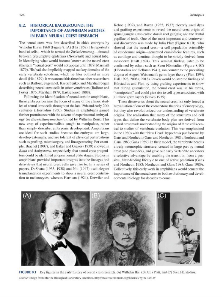
Wilhelm His in 1868 ( Figure 8.1A ) ( His 1868 ). He reported a
band of cells—which he termed the Zwischenstrang—situated
between presumptive epidermis (Hornblatt) and neural tube.
In identifying what would become known as the neural crest
(the term “neural crest” would not appear until 1879; Marshall
1879 ), His had also implicitly mapped out key domains of the
early vertebrate ectoderm, which he later outlined in more
detail ( His 1879 ). It was around this time that other researchers
such as Balfour, Sagemhel, Kastschenko, and Marshall began
describing neural crest cells in other vertebrates ( Balfour and
Foster 1876 ; Marshall 1879 ; Kastschenko 1888 ).
Following the identifcation of neural crest in amphibians,
these embryos became the focus of many of the classic studies of neural crest cells throughout the late 19th and early 20th
centuries ( Horstadius 1950 ). Studies in amphibians gained
further prominence with the advent of experimental embryology (or Entwicklungsmechanic), led by Wilhelm Roux. This
new crop of experimentalists sought to manipulate, rather
than simply describe, embryonic development. Amphibians
are ideal for such studies because the embryos are large,
develop externally, and are tolerant of physical perturbations
such as grafting, microsurgery, and lineage tracing. For example, Brachet (1907 ), and Baker and Graves (1939 ) showed in
Rana and Ambystoma, respectively, that neural crest progenitors could be identifed at open neural plate stages. Studies in
amphibians provided important insights into the lineages and
derivatives that neural crest cells give rise to. In a series of
papers, DuShane (1935 , 1938 ) and Niu (1947 ) used elegant
transplantation experiments to show a neural crest contribution to melanocytes, whereas Harrison (1924 ), Detwiler and
Kehoe (1939 ), and Raven (1935 , 1937 ) cleverly used dyes
and grafting experiments to reveal the neural crest origin of
spinal ganglia (also called dorsal root ganglia) and the dental
papillae of teeth. One of the most important and controversial discoveries was made by Julia Platt ( Figure 8.1B ), who
showed that the neural crest—a cell population ostensibly
of ectodermal origin—generated craniofacial features, such
as cartilage and dentine, thought to be strictly derived from
mesoderm ( Platt 1894 ). This seminal fnding, later to be
confrmed by others such as Sven Hörstadius ( Figure 8.1C )
( Hörstadius and Sellman 1941 ), ran counter to the prevailing
dogma of August Weissman’s germ layer theory ( Platt 1894 ;
Hall 1998 , 2008a, 2018 ). Raven would bolster the f ndings of
Hörstadius and Platt by using grafting experiments to show
that during gastrulation, the neural crest was, in his terms,
“omnipotent” and could give rise to cell types associated with
all three germ layers ( Raven 1935 ).
These discoveries about the neural crest not only forced a
reevaluation of one of the cornerstone theories of embryology,
but they also revolutionized our understanding of vertebrate
origins. The realization that many of the structures and cell
types that defne the vertebrate body plan are derived from
neural crest made understanding the origins of these cells central to studies of vertebrate evolution. This was emphasized
in the 1980s with the “New Head” hypothesis put forward by
Gans and Northcutt ( Gans and Northcutt 1983 ; Northcutt and
Gans 1983 ; Gans 1989 ). In their model, the vertebrate head is
a truly neomorphic structure, created in large part by neural
crest (and placodes), and gave our early vertebrate ancestors
a selective advantage by enabling the transition from a passive, flter-feeding lifestyle to one of active predation ( Gans
and Northcutt 1983 ; Northcutt and Gans 1983 ; Gans 1989 ).
Collectively, this early work in amphibians would cement the
importance of the neural crest to both evolutionary and developmental biology for decades to come.
FIGURE 8.1 Key fgures in the early history of neural crest research. (A) Wilhelm His, (B) Julia Platt, and (C) Sven Hörstadius.
Source: Image from Marine Biological Laboratory Archives, http://creativecommons.org/licenses/by-nc-sa/3.0/
Xenopus
8.2. HISTORICAL BACKGROUND: THE
IMPORTANCE OF AMPHIBIAN MODELS
IN EARLY NEURAL CREST RESEARCH
The neural crest was frst described in chick embryos by
Wilhelm His in 1868 ( Figure 8.1A ) ( His 1868 ). He reported a
band of cells—which he termed the Zwischenstrang—situated
between presumptive epidermis (Hornblatt) and neural tube.
In identifying what would become known as the neural crest
(the term “neural crest” would not appear until 1879; Marshall
1879 ), His had also implicitly mapped out key domains of the
early vertebrate ectoderm, which he later outlined in more
detail ( His 1879 ). It was around this time that other researchers
such as Balfour, Sagemhel, Kastschenko, and Marshall began
describing neural crest cells in other vertebrates ( Balfour and
Foster 1876 ; Marshall 1879 ; Kastschenko 1888 ).
Following the identifcation of neural crest in amphibians,
these embryos became the focus of many of the classic studies of neural crest cells throughout the late 19th and early 20th
centuries ( Horstadius 1950 ). Studies in amphibians gained
further prominence with the advent of experimental embryology (or Entwicklungsmechanic), led by Wilhelm Roux. This
new crop of experimentalists sought to manipulate, rather
than simply describe, embryonic development. Amphibians
are ideal for such studies because the embryos are large,
develop externally, and are tolerant of physical perturbations
such as grafting, microsurgery, and lineage tracing. For example, Brachet (1907 ), and Baker and Graves (1939 ) showed in
Rana and Ambystoma, respectively, that neural crest progenitors could be identifed at open neural plate stages. Studies in
amphibians provided important insights into the lineages and
derivatives that neural crest cells give rise to. In a series of
papers, DuShane (1935 , 1938 ) and Niu (1947 ) used elegant
transplantation experiments to show a neural crest contribution to melanocytes, whereas Harrison (1924 ), Detwiler and
Kehoe (1939 ), and Raven (1935 , 1937 ) cleverly used dyes
and grafting experiments to reveal the neural crest origin of
spinal ganglia (also called dorsal root ganglia) and the dental
papillae of teeth. One of the most important and controversial discoveries was made by Julia Platt ( Figure 8.1B ), who
showed that the neural crest—a cell population ostensibly
of ectodermal origin—generated craniofacial features, such
as cartilage and dentine, thought to be strictly derived from
mesoderm ( Platt 1894 ). This seminal fnding, later to be
confrmed by others such as Sven Hörstadius ( Figure 8.1C )
( Hörstadius and Sellman 1941 ), ran counter to the prevailing
dogma of August Weissman’s germ layer theory ( Platt 1894 ;
Hall 1998 , 2008a, 2018 ). Raven would bolster the f ndings of
Hörstadius and Platt by using grafting experiments to show
that during gastrulation, the neural crest was, in his terms,
“omnipotent” and could give rise to cell types associated with
all three germ layers ( Raven 1935 ).
These discoveries about the neural crest not only forced a
reevaluation of one of the cornerstone theories of embryology,
but they also revolutionized our understanding of vertebrate
origins. The realization that many of the structures and cell
types that defne the vertebrate body plan are derived from
neural crest made understanding the origins of these cells central to studies of vertebrate evolution. This was emphasized
in the 1980s with the “New Head” hypothesis put forward by
Gans and Northcutt ( Gans and Northcutt 1983 ; Northcutt and
Gans 1983 ; Gans 1989 ). In their model, the vertebrate head is
a truly neomorphic structure, created in large part by neural
crest (and placodes), and gave our early vertebrate ancestors
a selective advantage by enabling the transition from a passive, flter-feeding lifestyle to one of active predation ( Gans
and Northcutt 1983 ; Northcutt and Gans 1983 ; Gans 1989 ).
Collectively, this early work in amphibians would cement the
importance of the neural crest to both evolutionary and developmental biology for decades to come.
FIGURE 8.1 Key fgures in the early history of neural crest research. (A) Wilhelm His, (B) Julia Platt, and (C) Sven Hörstadius.
Source: Image from Marine Biological Laboratory Archives, http://creativecommons.org/licenses/by-nc-sa/3.0/